Leica Microsystems stellt die nächste Generation ihrer Konfokalplattform STELLARIS mit SpectraPlex vor.

Den Kunden in den Mittelpunkt zu stellen, Partnerschaften aufzubauen und Veränderungen im Sinne einer kontinuierlichen Verbesserung zu gestalten, wird…
Patentierte Lichtblattlösung von Viventis ermöglicht detaillierte volumetrische Bildgebung zur ganzheitlichen Erforschung des Lebens
Mehr sehen mit verbesserter Auflösung und reduzierter Lichtdosis
Bis zu 46% bessere Soma-Erkennung durch modernste KI und zusätzliche Funktionen für alle Forscher in der neuen Aivia-Version

Neue Ivesta 3 Stereomikroskope und Flexacam Kameras erweitern das Portfolio auf Basis der Enersight Softwareplattform.
Autonomous Microscopy powered by Aivia extrahiert maßgebliche Daten aus Experimenten und ermöglicht Wissenschaftlern so mehr zu entdecken
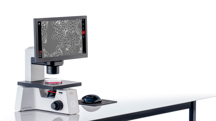
Problemlose Zellkontrolle mit dem Digitalmikroskop Mateo TL von Leica Microsystems

Vorstellung eines Einzelobjektiv-Lichtblattmikroskops auf der Cell Bio 2022

Einheitliche Benutzeroberfläche für QC-Community macht Inspektion mobil
