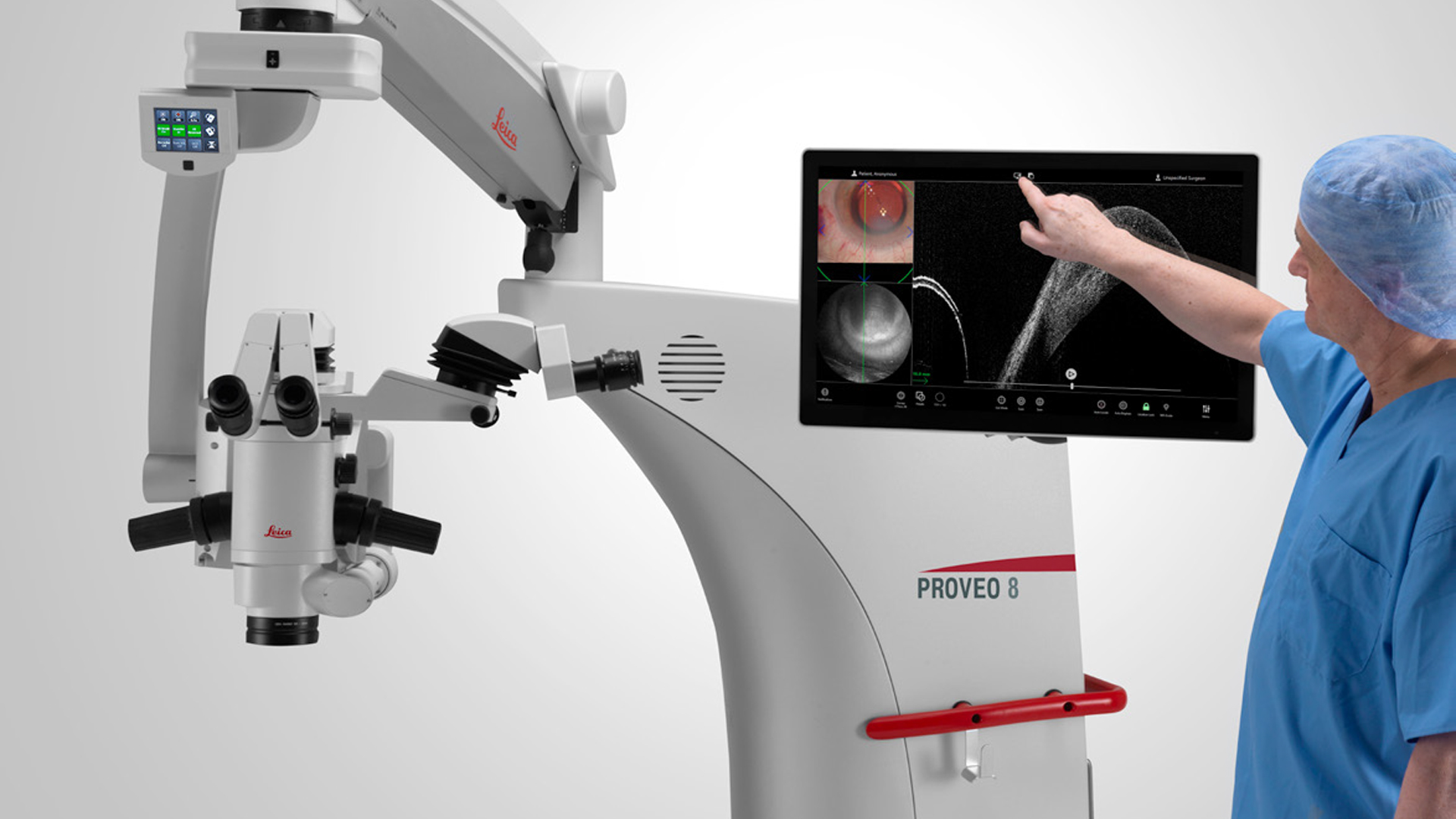
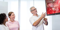

Das Proveo 8x 3D-Digitalmikroskop für die Ophthalmologie erlebt die Premiere auf der ASCRS 2025 in Los Angeles.
Medizinische Fachgebiete
Medizinische Fachgebiete
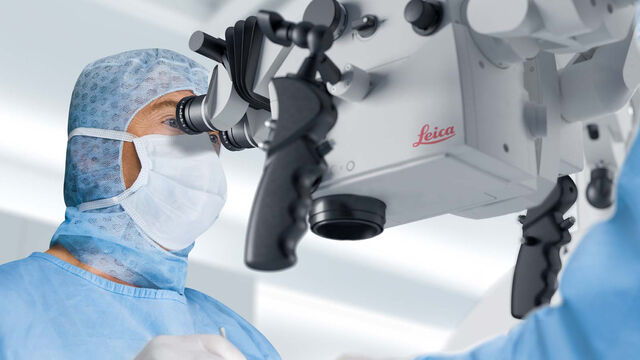
In medizinischen Anwendungen unterstützt Sie Leica mit hochwertigen Optiken, die eine klare, helle und konsistente Visualisierung ermöglichen. Um Ihre Sicht weiter zu verbessern, sind unsere hochwertigen Neurochirurgie- und Ophthalmologie-Mikroskope auch als erweiterbare Bildgebungsplattformen konzipiert. Integrieren Sie jederzeit zusätzliche digitale Bildgebungs- und Aufzeichnungstechnologien und stellen Sie sicher, dass Sie die Visualisierung haben, die Sie heute und morgen benötigen.
In medizinischen Anwendungen unterstützt Sie Leica mit hochwertigen Optiken, die eine klare, helle und konsistente Visualisierung ermöglichen.
Um Ihre Sicht weiter zu verbessern, sind unsere hochwertigen Neurochirurgie- und Ophthalmologie-Mikroskope auch als erweiterbare Bildgebungsplattformen konzipiert. Integrieren Sie jederzeit zusätzliche digitale Bildgebungs- und Aufzeichnungstechnologien und stellen Sie sicher, dass Sie die Visualisierung haben, die Sie heute und morgen benötigen.
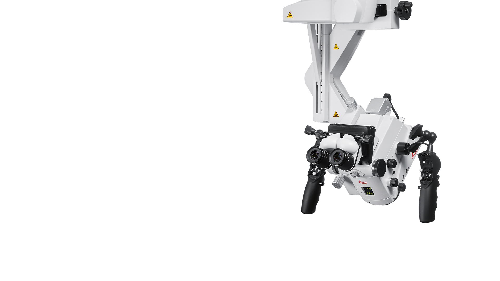
Mit einem Portfolio, das mikrochirurgische Disziplinen wie Ophthalmologie, Neurochirurgie, HNO, plastisch-rekonstruktive Chirurgie und Zahnmedizin umfasst, gibt es immer ein Operationsmikroskop von Leica für Ihre Bedürfnisse.

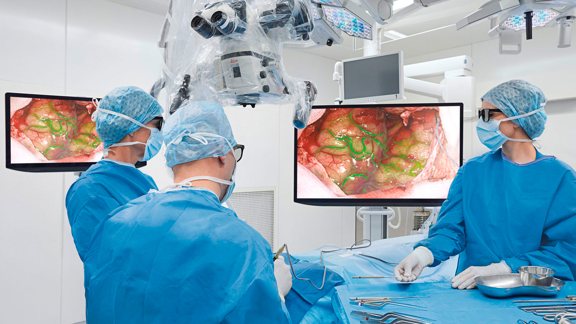
Jüngste Erweiterung des Operationsmikroskops ARveo 8 schafft klinischen Mehrwert bei der 3D-Visualisierung von Hirntumoren und ermöglicht neue…

Interview über den Einzug von Operationsmikroskopen im HNO-Bereich
Werfen Sie einen Blick auf unsere kommenden Kongresse, Ausstellungen, Webinare und Workshops und besuchen Sie uns bei einer unserer nächsten Veranstaltungen!
25
–
27
Mar
2026
Congrès SFNC 2026
France
•
Congress
09
–
11
May
2026
Congrès SFO 2026
France
•
Congress
