
Andrea Boni , PhD

Andrea Boni studierte Biotechnologie an der Universität San Raffaele in Mailand, bevor er für seine Promotion an das EMBL-Labor in Heidelberg (Deutschland) wechselte, wo er sich mit dem mikroskopischen Screening der Dynamik von Membran-Kernproteinen befasste. Anschließend wechselte er zur Entwicklung von Lichtblättern und zur Bildgebung von Darmorganoiden. 2017 war Andrea Mitbegründer von Viventis Microscopy, wo er für die Bildgebungsanwendungen und den Vertrieb verantwortlich war. Nach der Integration von Viventis Microscopy in Leica Microsystems kam er im Mai 2024 als Business Excellence Director zu Leica.
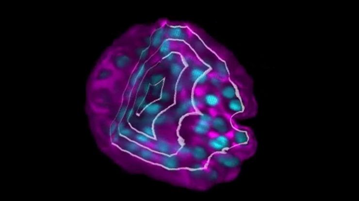
Advancing ADC Development with Imaging of 3D Tumor Models
Antibody-drug conjugates (ADCs) are targeted therapies designed to selectively deliver drugs to cancer cells. Evaluating ADC behavior in 3D tumor models more closely reflects the physiological…
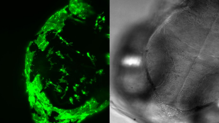
Capturing Developmental Dynamics in 3D
This application note showcases how the Viventis Deep dual-view light sheet microscope was successfully used by researchers for exploring high-resolution, long-term imaging of 3D multicellular models…


