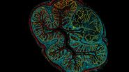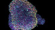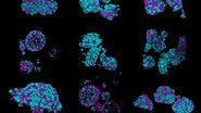
How to optimize the biological relevance of 3D models
ADC success starts with a biologically relevant and reproducible 3D model. Researchers need to verify that their 3D tumor models consistently express the target antigen, remain viable over time, and accurately reflect disease biology before progressing to ADC evaluation. Early characterization is critical for selecting appropriate models, confirming assay suitability, identifying relevant biomarkers, and reducing development risk.
How Leica can help
- The Mateo FL Digital Microscope supports routine monitoring of 3D cultures directly in the cell culture hood with quick, reproducible, label-free imaging. This enables assessment of growth, morphology, and culture consistency without disrupting experiments.
- For deeper biological insight, the Viventis Deep Dual View Light Sheet Microscope enables visualization of receptor distribution and target expression throughout intact 3D models, helping researchers understand spatial heterogeneity and confirm target accessibility.
Our Solution
Viventis Deep Dual View Light Sheet Microscope
Go beyond "is my ADC target expressed?" to "where and how much is it expressed throughout my 3D tumor model?"
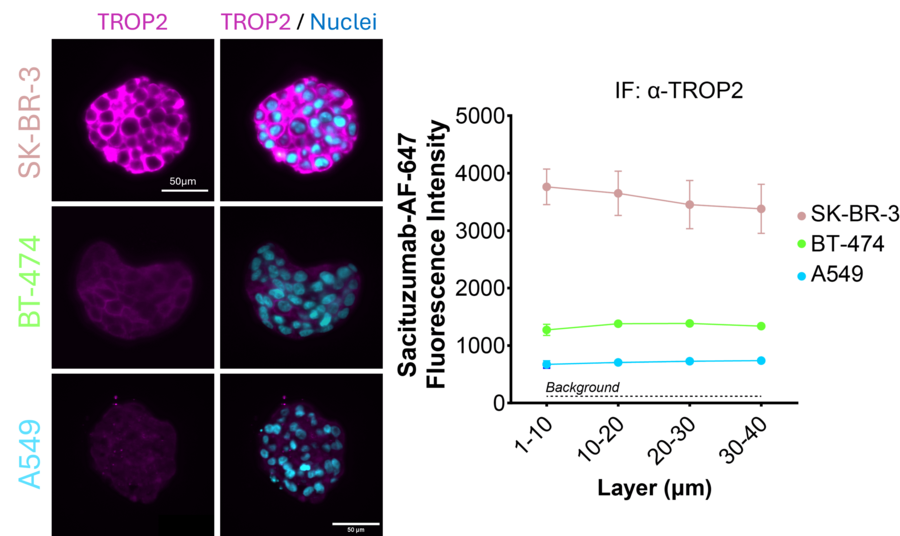
TROP2 target expression across different 3D tumor models characterized by light-sheet fluorescence imaging. Representative images show anti-TROP2 immunofluorescence (magenta) and nuclei SPY-DNA555 (cyan) in SK-BR-3 and BT-474 breast 3D tumor models, and A549 3D lung tumor model. These were generated using FluoSphera’s cell encapsulation technology to produce size-controlled tumor spheroids. Depth-resolved quantification demonstrates strong TROP2 expression in SK-BR-3, moderate expression in BT-474, and ultra-low expression in A549. Viventis Deep enables volumetric assessment of target expression across successive layers of the 3D models, providing information on the spatial distribution of the ADC target throughout the sample.
How to measure ADC Uptake, Penetration and Intracellular Trafficking
To achieve a therapeutic effect, the ADC must first penetrate throughout the tumor to relevant areas and, after binding to the target antigen, enter the cell and release its payload. Advanced imaging allows researchers to directly visualize ADC distribution in 3D tumor models, distinguish surface-bound from internalized ADCs, track intracellular localization, and monitor payload release.
How Leica can help
- Viventis Deep light sheet microscopy enables imaging of intact tumor models to quantify ADC penetration and spatial distribution.
- STELLARIS delivers high-resolution live-cell imaging to study internalization, intracellular trafficking, and subcellular localization.
Our Solution
Viventis Deep Dual View Light Sheet Microscope
Reveal how fast and how deep your ADC penetrates a 3D tumor model
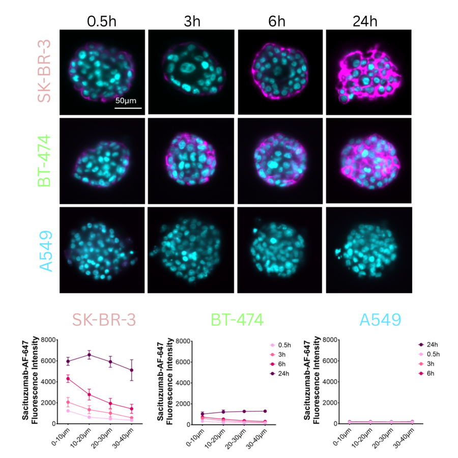
Time-dependent antibody (Sacituzumab) penetration in 3D solid tumor models. SK-BR-3, BT-474, and A549 3D tumor models were imaged at 0.5h, 3h, 6h, and 24h after exposure. The unconjugated antibody is labelled with a fluorescence dye AF-647 (magenta) using Debiopharm’s AbYlink™ conjugation technology, enabling rapid one-step Fc directed conjugation (Licensed by FluoSphera). In SK-BR-3 and BT-474, intensity increases over time, progressing from peripheral regions toward deeper areas of the 3D models. In A549, which only poorly express the antibody target, minimal signal is observed. Viventis Deep combines dual-sided illumination and dual-view detection to support deep volumetric imaging and maintain image quality across complex, light-scattering 3D samples. This approach enables spatial assessment of antibody penetration throughout the 3D cancer model, as demonstrated here, and is applicable to fluorescently labelled ADCs. Nuclei are shown in cyan. Scale bar; 50 µm. Courtesy of Simon Latour, FluoSphera SA.
Our Solution
STELLARIS Confocal Microscope Platform
Go beyond ADC penetration: reveal where your ADC travels inside tumor cells
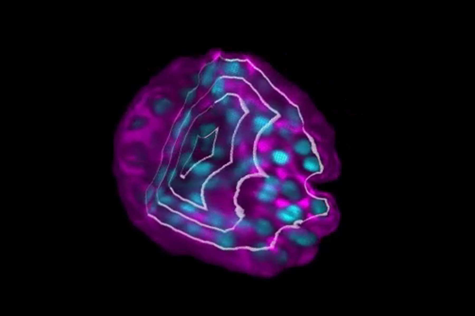
Tracking ADC delivery to lysosomes is particularly relevant. This is because lysosomal processing can promote linker cleavage and/or antibody degradation, enabling intracellular payload release and connecting target engagement with the ADC's cytotoxic mechanism of action. High-resolution confocal imaging with STELLARIS can provide the contrast and spatial detail required to visualize ADC uptake and its intracellular localization relative to lysosomal compartments. The movie below shows vesicle transport imaged with Fluorescence Lifetime Imaging (FLIM) on STELLARIS FALCON. As pH decreases, the fluorescence lifetime shortens, allowing trafficking to be followed from early endosomes (cyan/yellow) through late endosomes to lysosomes (magenta/red).
How to quantify the tumor killing efficacy of ADCs
Once an ADC reaches tumor cells, the next question is whether it produces the intended therapeutic effect. Researchers need to understand which cell populations are killed, which survive, and whether resistant subpopulations emerge. In complex and heterogeneous 3D tumor models, this can be challenging to measure. Quantitative imaging helps reveal how response changes over time, providing the evidence needed to compare candidates, understand mechanisms of action, and make confident development decisions.
How Leica can help
- The Mica Microhub enables automated imaging in multi-well plates, allowing 3D tumor viability to be assessed using morphological features and fluorescence reporters.
- Mica and Aivia allow scientists to rapidly unlock AI-powered insights from multi-well plate assays, without coding or labour-intensive deep learning training. The results can be visualized in interactive charts such as heatmaps and dose response curves.
Acknowledgement
The authors gratefully acknowledge FluoSphera for proving the encapsulated 3D cancer models and generating the data, and Debiopharm Research & Manufacturing SA for kindly providing Sacituzumab-AF-647.
FAQs
Why are 3D tumor models more relevant than 2D cultures for studying tissue penetration?
3D tumor models, such as organoids and spheroids, reproduce key features of solid tumors, including cell-cell interactions, extracellular matrix, and oxygen and nutrient gradients. These features create realistic barriers to drug penetration, making them more representative of in vivo conditions.
How can I determine whether my target biomarker is expressed at sufficient levels in a 3D tumor model?
Assessing target biomarker expression is a critical first step in ADC development. Imaging can help visualize where the biomarker is expressed, quantify expression levels, and reveal heterogeneity across different regions and cell populations within a 3D tumor model. These insights help researchers evaluate whether a target is suitable for ADC-mediated delivery.
How deeply does an ADC penetrate complex 3D tumor models?
ADC penetration is an important determinant of therapeutic efficacy. Imaging can visualize the spatial distribution of ADCs throughout a 3D tumor model and help assess whether the therapeutic reaches cells beyond the outer layers of the tissue construct.
What factors influence ADC distribution within a tumor model?
ADC distribution can be affected by target expression, tissue architecture, cellular density, and other biological characteristics of the model. Imaging provides spatial information that helps researchers understand how these factors influence ADC localization and movement throughout the tissue.
Is my ADC efficiently internalized by target cells after binding?
Effective ADC activity depends on cellular uptake following target binding. Imaging enables researchers to monitor ADC localization over time and distinguish surface-bound ADCs from internalized molecules. This helps evaluate whether the ADC reaches intracellular compartments required for payload release.
How efficiently does an ADC eliminate target cells while minimizing off-target effects?
Evaluating efficacy requires understanding both target cell response and potential effects on non-target cells. Imaging can support the assessment of cellular viability, morphology, and spatial response patterns within complex tumor models, helping researchers characterize treatment outcomes.