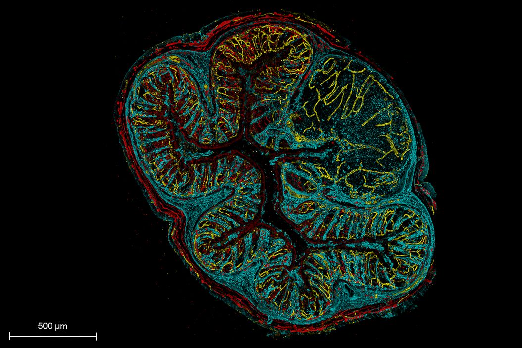
Introduction
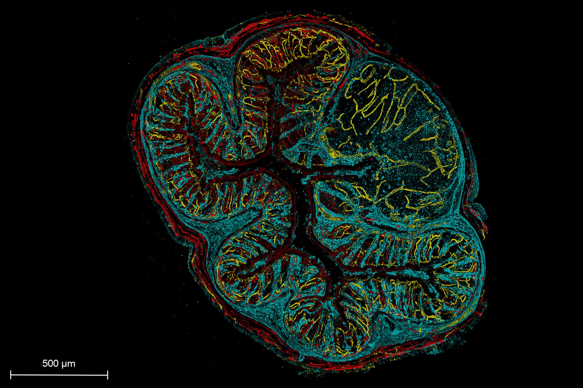
Studying biological processes in three dimensions requires imaging methods that combine volumetric speed with controlled light exposure. This webinar explores the principles of oblique plane microscopy and its application to imaging cells, organoids, tissues and cleared specimens, with examples illustrating biological dynamics and structural changes across space and time.
From Fast Dynamics to Scalable 3D Experiments
Capture biological processes in three dimensions
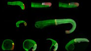
Fast biological events can be difficult to interpret when they are recorded in only one focal plane. Rapid volumetric imaging captures information across depth, helping researchers follow movement, signaling and structural changes in their full spatial context. The webinar includes examples involving calcium transients, cell morphology, biosensor activity, tissue dynamics and small model organisms.
Preserve the biology during live imaging
Illuminating only the plane being recorded reduces unnecessary exposure outside the focal region. This is especially relevant for light-sensitive samples, repeated volumetric acquisition and long-term time-lapse experiments.
Scale imaging across samples and conditions
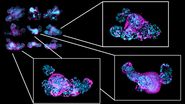
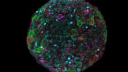
Fast volume acquisition can increase the number of samples, positions and time points included in an experiment. The organoid pilot study demonstrates volumetric imaging across different organoid lines and extended live-imaging experiments, supporting the development of scalable 3D screening workflows.
Work with familiar sample formats
The single-objective inverted configuration supports conventional flat-bottom carriers, including microscope slides, glass-bottom dishes and multiwell plates. This reduces dependence on specialized mounting and helps integrate light sheet imaging into established experimental workflows.