Specialità mediche
Specialità mediche
Esplora una raccolta completa di risorse scientifiche e cliniche su misura per gli operatori sanitari, tra cui approfondimenti tra pari, casi clinici e simposi. Progettata per neurochirurghi, oculisti e specialisti in chirurgia plastica e ricostruttiva, otorinolaringoiatria e odontoiatria. Questa raccolta evidenzia gli ultimi progressi nel campo della microscopia chirurgica. Scoprite come le tecnologie chirurgiche all'avanguardia, come la fluorescenza AR, la visualizzazione 3D e l'imaging OCT intraoperatorio, consentano di prendere decisioni sicure e di essere precisi in interventi chirurgici complessi.
Artificial Intelligence and Confocal Microscopy – What You Need to Know
This list of frequently asked questions provides “hands-on” answers and is a supplement to the introductory article about Dynamic Signal Enhancement powered by Aivia "How Artificial Intelligence…
How Artificial Intelligence Enhances Confocal Imaging
In this article, we show how artificial intelligence (AI) can enhance your imaging experiments. Namely, how Dynamic Signal Enhancement powered by Aivia improves image quality while capturing the…
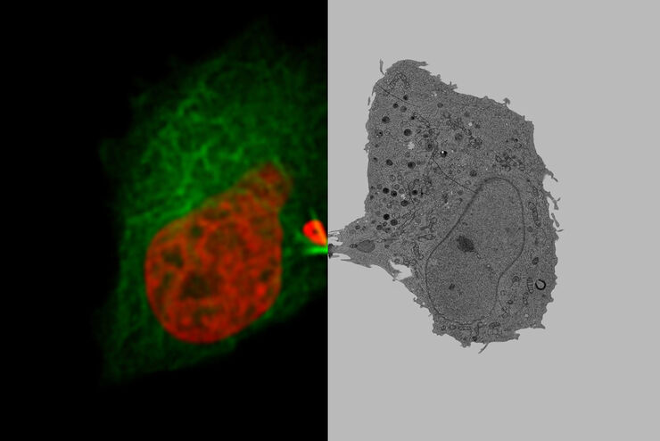
How to Successfully Perform Live-Cell CLEM
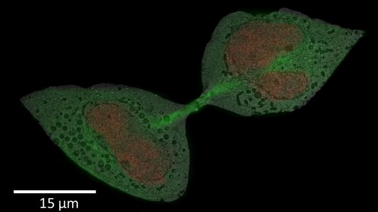
The Leica Nano workflow provides a streamlined live-cell CLEM solution for getting insight bout structural changes of cellular components over time. Besides the technical handling described in the…
How to Improve Live Cell Imaging with Coral Life
For live-cell CLEM applications, light microscopy imaging is a critical step for identifying the right cell in the right state at the right time. In this article, Leica experts share their insights on…
How to Keep Your Samples Under Physiological Conditions
The Coral Life workflow combines dynamic data with the best possible sample fixation by high pressure freezing. However, good sample preservation won’t help if your cells are stressed by temperature…
Physiology Image Gallery
Physiology is about the processes and functions within a living organism. Research in physiology focuses on the activities and functions of an organism’s organs, tissues, or cells, including the…
Live Cell Imaging Gallery
Live cell microscopy techniques are fundamental to get a better understanding of cellular and molecular function. Today, widefield microscopy is the most common technique used to visualize cell…
Microscopi per campo scuro
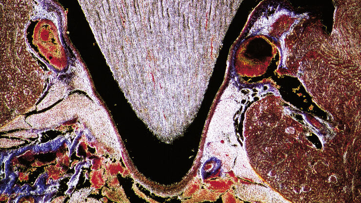
Il metodo di contrasto in campo oscuro sfrutta la diffrazione o la dispersione della luce da strutture di un campione biologico o da caratteristiche non uniformi nella struttura di un materiale.
Putting Dynamic Live Cell Data into the Ultrastructural Context
With workflow Coral Life, searching for a needle in the haystack is a thing of the past. Take advantage of correlative light and electron microscopy to identify directly the right cell at the right…

