STELLARIS FALCON
共焦点顕微鏡
製品紹介
Home
Leica Microsystems
STELLARIS FALCON 蛍光寿命イメージングを素早く入手
クリアなコントラスト
最新の記事を読む
 and acceptor (A) molecule which participate in FRET (Förster resonance energy transfer).")
What is FRET with FLIM or as it is usually known FLIM-FRET?
Förster resonance energy transfer (FRET) is a well-established fluorescence-based technique which is used to study molecular interactions. It is useful for the analysis of protein-DNA and…

Researchers Insights: Microscopy in Cancer Research
Discover how imaging techniques are driving cancer research forward. In this issue, we present comprehensive multimodal studies using microscopy, as well as new directions in intraoperative cancer…
, Tropomyosin (cardiomyocytes, red) and GFP (primordial cardiac layer, green).")
A Guide to Fluorescence Microscopy
Fluorescence microscopy uses the ability of fluorophores, dyes, or fluorescent proteins to emit light of a specific wavelength after being excited with light of a shorter wavelength. Biomolecules can…
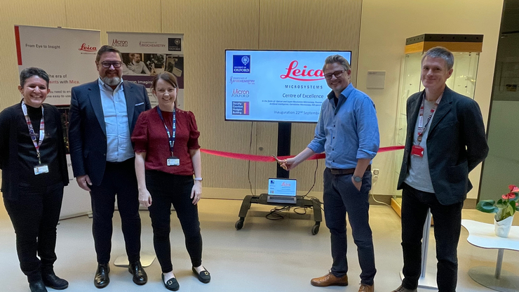
Micron Bioimaging Facility at the University of Oxford
Find out how to access cutting-edge microscopy systems, with dedicated training and support, at the Micron Bioimaging Facility at the University of Oxford’s Department of Biochemistry.
. Courtesy: Thomas Mathivet, PhD")
Windows on Neurovascular Pathologies
Discover how innate immunity can sustain deleterious effects following neurovascular pathologies and the technological developments enabling longitudinal studies into these events.

Live-Cell Fluorescence Lifetime Multiplexing Using Organic Fluorophores
On-demand video: Imaging more subcellular targets by using fluorescence lifetime multiplexing combined with spectrally resolved detection.

Visualizing Protein-Protein Interactions by Non-Fitting and Easy FRET-FLIM Approaches
The Webinar with Dr. Sergi Padilla-Parra is about visualizing protein-protein interaction. He gives insight into non-fitting and easy FRET-FLIM approaches.
 in 14 x 18 tiles. Lifetime gives an additional contrast that allows to differentiate different structures in histological stainings.")
A Guide to Fluorescence Lifetime Imaging Microscopy (FLIM)
The fluorescence lifetime is a measure of how long a fluorophore remains on average in its excited state before returning to the ground state by emitting a fluorescence photon.

Find Relevant Specimen Details from Overviews
Switch from searching image by image to seeing the full overview of samples quickly and identifying the important specimen details instantly with confocal microscopy. Use that knowledge to set up…

Fluorescence Lifetime-based Imaging Gallery
Confocal microscopy relies on the effective excitation of fluorescence probes and the efficient collection of photons emitted from the fluorescence process. One aspect of fluorescence is the emission…

How to Quantify Changes in the Metabolic Status of Single Cells
Metabolic imaging based on fluorescence lifetime provides insights into the metabolic dynamics of cells, but its use has been limited as expertise in advanced microscopy techniques was needed.
Now,…

How FLIM Microscopy Helps to Detect Microplastic Pollution
The use of autofluorescence in biological samples is a widely used method to gain detailed knowledge about systems or organisms. This property is not only found in biological systems, but also…

LIGHTNINGによって試料から最大限の情報を引き出す
LIGHTNINGは、他の方法では簡単に可視化できない、微細な構造や形態を完全自動で明らかにする、適応能力に優れた情報抽出プロセスです。 LIGHTNINGは、画像全体を同一のパラメーターで演算する従来型の手法とは異なり、ボクセル(3次元画素)ごとに適切なパラメーターを算出することによって、最高の忠実度であらゆる微細形態を明らかにします。

ウイルス学
ウイルス研究のためのイメージングと試料作製ソリューション

Microscopy in Virology
The coronavirus SARS-CoV-2, causing the Covid-19 disease effects our world in all aspects. Research to find immunization and treatment methods, in other words to fight this virus, gained highest…

TauSense Technology Imaging Tools
Leica Microsystems’ TauSense technology is a set of imaging modes based on fluorescence lifetime. Found at the core of the STELLARIS confocal platform, it will revolutionize your imaging experiments.…
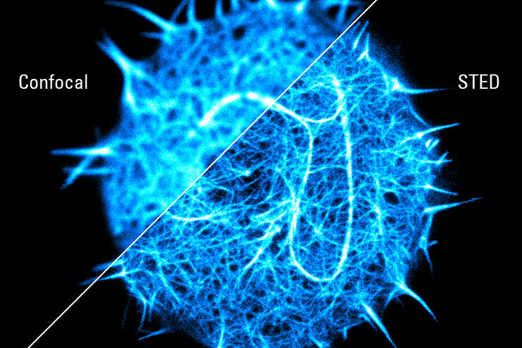
Super-resolved STED spectroscopy
Molecular interactions are key in cellular signalling. They are often ruled or rendered by the mobility of the involved molecules.

Förster Resonance Energy Transfer (FRET)
The Förster Resonance Energy Transfer (FRET) phenomenon offers techniques that allow studies of interactions in dimensions below the optical resolution limit. FRET describes the transfer of the energy…
Fields of Application
高度な組織イメージングおよび解析
Leica Microsystems の高度なイメージングソリューションを活用することで、組織の構造および機能に対する理解を深め、空間生物学や疾患機構の解明を促進します。
