Windows on Neurovascular Pathologies
Webinar on-demand
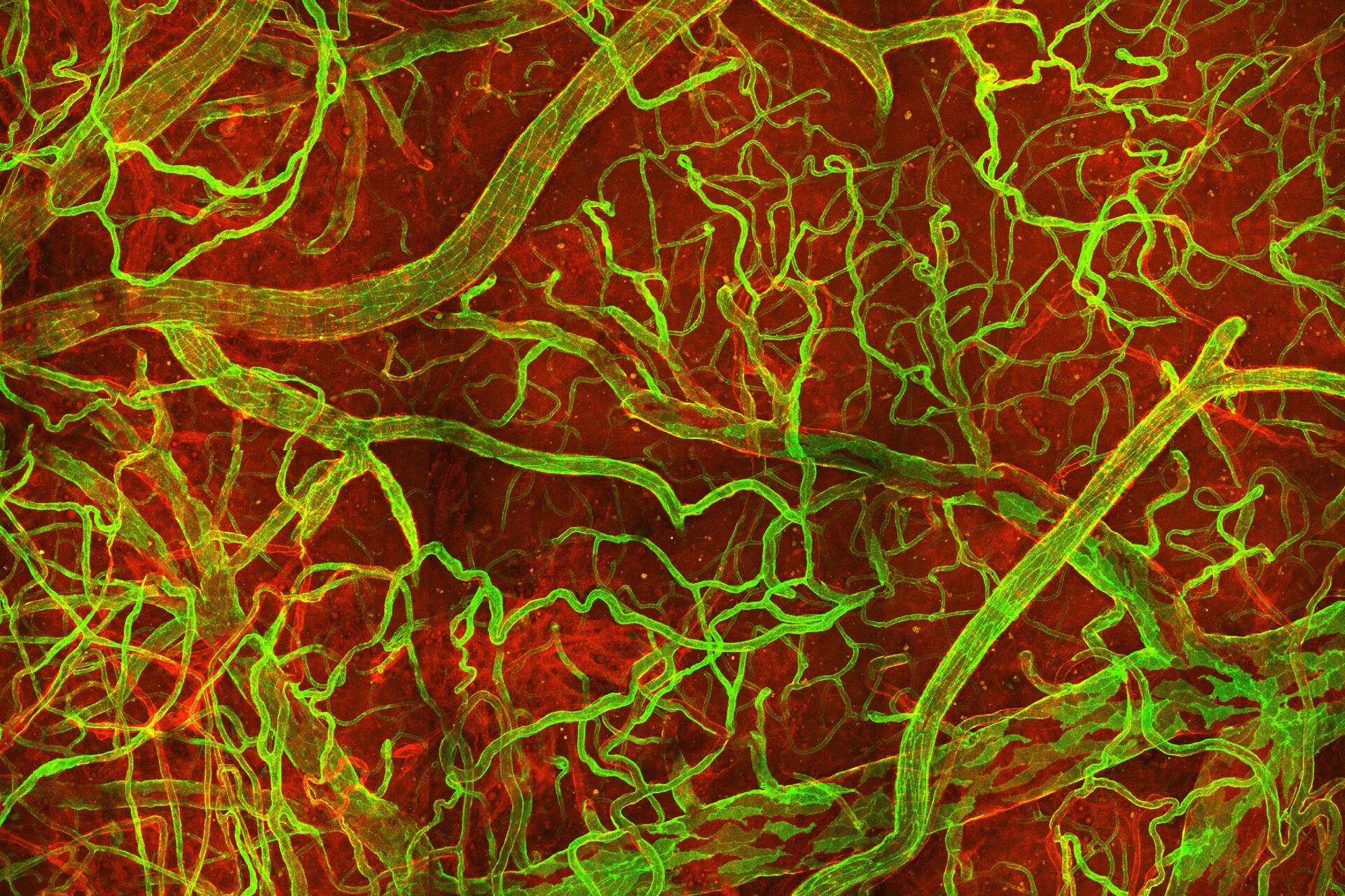
. Courtesy: Thomas Mathivet, PhD")
Summary
The brain is an immune-privileged organ, meaning it is protected from the entry of pathogens, blood-circulating factors, and immune cells by a physical barrier called the blood–brain barrier (BBB). But pathological events, such as ischemia (stroke), trauma (traumatic brain injury), or tumor growth (gliomas and brain metastasis), activate the vascular endothelium, disturb or disrupt the BBB, and allow immune cell homing.
In this context, innate immunity, which is recruited at the lesion site to prevent neuro-vascular damages, is rewired by the pathological microenvironment and ends up sustaining deleterious effects such as edema formation, perfusion defects, and hemorrhages.
Such analysis of dynamical processes, cell mobility, cell-cell interactions, and implicated signaling pathways in neurovascular pathologies was facilitated by genetic tool developments, such as inducible fluorescent mice lineage, and novel technologies of longitudinal imaging compatible with in vivo work, such as multiphoton microscopy.
Discover the dynamic remodeling of the microenvironment during neurovascular pathologies, the plasticity of innate immunity and which phenotypes switch in such microenvironments, and the technologies that enabled us to unravel these events longitudinally.
After the presentation, join Ulf Schwarz for a live showcase demonstration of the Leica STELLARIS 8 DIVE Multicolor Multiphoton Microscope. Experience a rainbow of possibilities to expand your research. At the heart of STELLARIS 8 DIVE is 4Tune, the first and only spectral non-descanned detection system.
The showcase will demonstrate how easily experimental settings are defined for multicolor multiphoton imaging with STELLARIS 8 DIVE and how you can benefit from lifetime-based information using TauSense or Fast Lifetime Contrast (FALCON).
Key learnings
- The dynamic remodeling of the microenvironment during neurovascular pathologies;
- The plasticity of our innate immunity and how it sustains deleterious effects;
- Technological developments that are enabling longitudinal studies of mice central nervous system
Discover how innate immunity can sustain deleterious effects following neurovascular pathologies and the technological developments enabling longitudinal studies into these events.
Related Articles
-
 and phalloidin (magenta), imaged using Viventis SCAPE; scale bar 50μm. Courtesy of Marina Cuenca and Heleen Jungen (Dayton lab), EMBL Barcelona.")
What’s the Best Organoid Imaging Approach for Early Drug Discovery?
Organoids and other complex in vitro models (CIVMs) are becoming increasingly important in early…
Jun 30, 2026Read article -
 and acceptor (A) molecule which participate in FRET (Förster resonance energy transfer).")
What is FRET with FLIM or as it is usually known FLIM-FRET?
Förster resonance energy transfer (FRET) is a well-established fluorescence-based technique which is…
Apr 21, 2026Read article -

History, Developments and Trends of Microscopy in Cancer Research
Cancer is a global disease, with 18 million new cases diagnosed and 10 million cancer-related deaths…
Mar 16, 2026Read article
