EM ICE
電顕用試料作製装置
製品紹介
Home
Leica Microsystems
EM ICE 高圧凍結装置
その」瞬間を凍結
最新の記事を読む
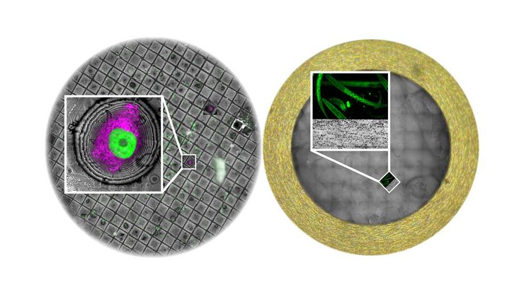
 labeled with membrane-permeable calcein, high-pressure frozen in salt water using EM ICE.")
High-Pressure Freezing for Organoids: Cryo CLEM & FIB Lift Out
Master cryo EM workflow steps for challenging 3D samples: when to choose HPF vs. plunge freezing, reproducible blotting/ice control, contamination aware transfers, Cryo CLEM 3D targeting in organoids,…
 image of a cross section of C. elegans (roundworm). Courtesy of T. Müller-Reichert, MPI-CBG, Dresden, Germany and K. McDonald, University of California, Berkeley, USA.")
Brief Introduction to High-Pressure Freezing for Cryo-Fixation
Preparation of biological specimens for electron microscopy (EM) often requires cryo-fixation which does not introduce significant structural alterations of cellular constituents. A common method used…

神経科学研究
神経変性疾患の理解向上に取り組んでいる、もしくは神経系の機能を研究をしていますか? ライカマイクロシステムズのイメージングソリューションによってブレイクスルーを起こす方法をご覧ください。

The “Waffle Method”: High-Pressure Freeze Complex Samples
This article describes the advantages of a special high pressure freezing method, the so-called “Waffle Method”. Learn how the “Waffle Method” uses EM grids as spacers for high-pressure freezing,…

How Marine Microorganism Analysis can be Improved with High-pressure Freezing
In this application example we showcase the use of EM-Sample preparation with high pressure freezing, freeze substiturion and ultramicrotomy for marine biology focusing on ultrastructural analysis of…

How to Successfully Perform Live-Cell CLEM
The Leica Nano workflow provides a streamlined live-cell CLEM solution for getting insight bout structural changes of cellular components over time. Besides the technical handling described in the…

How to Improve Live Cell Imaging with Coral Life
For live-cell CLEM applications, light microscopy imaging is a critical step for identifying the right cell in the right state at the right time. In this article, Leica experts share their insights on…

How to Keep Your Samples Under Physiological Conditions
The Coral Life workflow combines dynamic data with the best possible sample fixation by high pressure freezing. However, good sample preservation won’t help if your cells are stressed by temperature…

Putting Dynamic Live Cell Data into the Ultrastructural Context
With workflow Coral Life, searching for a needle in the haystack is a thing of the past. Take advantage of correlative light and electron microscopy to identify directly the right cell at the right…

Fast, High-quality Vitrification with the EM ICE High Pressure Freezer
The EM ICE High Pressure Freezer was developed with a unique freezing principle and uses only a single pressurization and cooling liquid: liquified nitrogen (LN2). This design enables three major…

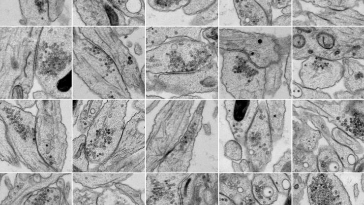
Investigating Synapses in Brain Slices with Enhanced Functional Electron Microscopy
A fundamental question of neuroscience is: what is the relationship between structural and functional properties of synapses? Over the last few decades, electrophysiology has shed light on synaptic…

High-pressure freezing: Revealing functional mechanisms of synaptic transmission
Learn more about applying optogenetic stimulation in the EM ICE and how this technology has the potential to reveal structural and functional mechanisms of synaptic transmission. Get a detailed…
Workflows and Instrumentation for Cryo-electron Microscopy
Cryo-electron microscopy is an increasingly popular modality to study the structures of macromolecular complexes and has enabled numerous new insights in cell biology. In recent years, cryo-electron…

研究におけるモデル生物
モデル生物とは、特定の生物学的プロセスを研究するために研究者が使用する生物種です。 モデル生物は、人間と似た遺伝的特徴を持ち、遺伝子学、発生生物学、神経科学などの研究分野で一般的に使用されています。 通常、モデル生物は実験環境での維持や繁殖が容易であること、生殖サイクルが短いこと、または、特定の形質や病気を研究するために突然変異体を生成する能力を持つことで選ばれます。

ウイルス学
ウイルス研究のためのイメージングと試料作製ソリューション

Expert Knowledge on High Pressure Freezing and Freeze Fracturing in the Cryo SEM Workflow
Get an insight in the working methods of the laboratory and learn about the advantages of Cryo SEM investigation in EM Sample Preparation. Find out how high pressure freezing, freeze fracturing and…
Bridging Structure and Dynamics at the Nanoscale through Optogenetics and Electrical Stimulation
Nanoscale ultrastructural information is typically obtained by means of static imaging of a fixed and processed specimen. However, this is only a snapshot of one moment within a dynamic system in…
EM Sample Prep Workflows & Uses
Researchers can consistently achieve high-quality, precise, and reproducible results when imaging samples with electron microscopy by using Leica sample preparation solutions. Our solutions support…
Fields of Application
光電子相関顕微鏡法(CLEM)
ライカマイクロシステムズの Coral ワークフローによって、蛍光顕微鏡と電子顕微鏡のデータを相関させることができます (CLEM)。
