
Industrial
Industrial
Mergulhe fundo em artigos detalhados e webinars com foco em inspeção eficiente, fluxos de trabalho otimizados e conforto ergonômico em contextos industriais e patológicos. Os tópicos abordados incluem controle de qualidade, análise de materiais, microscopia em patologia, entre muitos outros. Este é o lugar onde você obtém insights valiosos sobre o uso de tecnologias de ponta para melhorar a precisão e a eficiência dos processos de fabricação, bem como o diagnóstico e a pesquisa patológicos precisos.

Mission Impossible Accomplished: Tunable Colors for Non-descanning Detection
Leica Microsystems’ 4Tune detector, the key component of the SP8 DIVE Deep In Vivo Explorer, provides spectrally tunable image recording with non-descanning detection. An innovative solution for…

Introduction to Mammalian Cell Culture
Mammalian cell culture is one of the basic pillars of life sciences. Without the ability to grow cells in the lab, the fast progress in disciplines like cell biology, immunology, or cancer research…

Laser Beam Shaping for Multicolor Multiphoton Microscopy
Multiphoton Microscopy is one of the current hot topics in life science research. The new Leica TCS SP8 DIVE from Leica Microsystems presents a series of beneficial new innovations, including a freely…
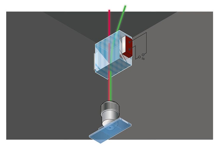
Primary Beam Splitting Devices for Confocal Microscopes
Current fluorescence microscopy employs incident illumination which requires separation of illumination and emission light. The classical device performing this separation is a color-dependent beam…

Photoactivatable, Photoconvertible, and Photoswitchable Fluorescent Proteins
Fluorescent proteins (FPs) such as GFP, YFP or DsRed are powerful tools to visualize cellular components in living cells. Nevertheless, there are circumstances when classical FPs reach their limits.…

Pinhole Effect in Confocal Microscopes
When operating a confocal microscope, or when discussing features and parameters of such a device, we inescapably mention the pinhole and its diameter. This short introductory document is meant to…
 stereo microscope for a task like surgery.")
Rodent and Small-Animal Surgery
Learn how you can perform rodent (mouse, rat, hamster) and small-animal surgery efficiently with a microscope for developmental biology and medical research applications by reading this article.
 illuminated with wide-band UV excitation. Note the tissue structure with blue autofluorescence. Right: Same tissue and same immunostaining with FITC label illuminated with epi-illumination using narrow")
Milestones in Incident Light Fluorescence Microscopy
Since the middle of the last century, fluorescence microscopy developed into a bio scientific tool with one of the biggest impacts on our understanding of life. Watching cells and proteins with the…

Chronic Inflammation Under the Microscope
In the course of chronic inflammation certain body areas are recurrently inflamed. This goes along with many human diseases. With the help of widefield light microscopy, the underlying processes can…
