STELLARIS STED
共焦点顕微鏡
製品紹介
Home
Leica Microsystems
STELLARIS STED
より速く真実にたどり着く
最新の記事を読む

Researchers Insights: Microscopy in Cancer Research
Discover how imaging techniques are driving cancer research forward. In this issue, we present comprehensive multimodal studies using microscopy, as well as new directions in intraoperative cancer…
, Tropomyosin (cardiomyocytes, red) and GFP (primordial cardiac layer, green).")
A Guide to Fluorescence Microscopy
Fluorescence microscopy uses the ability of fluorophores, dyes, or fluorescent proteins to emit light of a specific wavelength after being excited with light of a shorter wavelength. Biomolecules can…

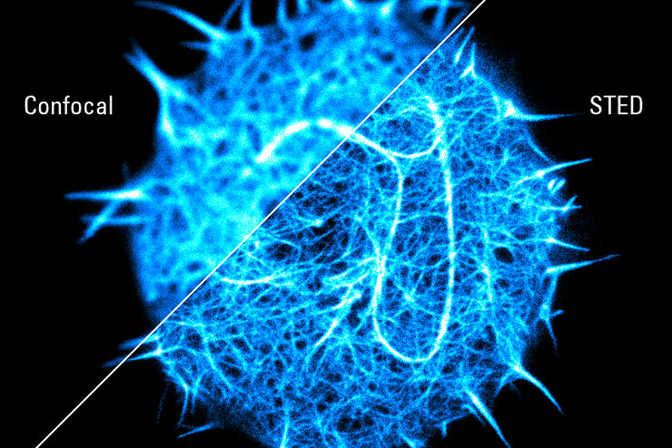
Super-Resolution Microscopy Image Gallery
Due to the diffraction limit of light, traditional confocal microscopy cannot resolve structures below ~240 nm. Super-resolution microscopy techniques, such as STED, PALM or STORM or some…

Extended Live-cell Imaging at Nanoscale Resolution
Extended live-cell imaging with TauSTED Xtend. Combined spatial and lifetime information allow super-resolution microscopy at extremely low light dose.
, actin network (ATTO 647N), and nuclear pore basket (CF 680R).")
The Guide to STED Sample Preparation
This guide is intended to help users optimize sample preparation for stimulated emission depletion (STED) nanoscopy, specifically when using the STED microscope from Leica Microsystems. It gives an…

Studying Virus Replication with Fluorescence Microscopy
The results from research on SARS-CoV-2 virus replication kinetics, adaption capabilities, and cytopathology in Vero E6 cells, done with the help of fluorescence microscopy, are described in this…

Five-color FLIM-STED with One Depletion Laser
Webinar on five-color STED with a single depletion laser and fluorescence lifetime phasor separation.

A Versatile Palette of Fluorescent Probes
Researchers at the Max Planck Institute for Medical Research in Heidelberg have developed a general strategy to synthesize live-cell compatible fluorogenic probes, and the result are the new MaP (Max…

Benefits of Combining STED and Lifetime
In this interview, Professor Alberto Diaspro talks about the advantages of the White Light Laser and the TauSTED capabilities of STELLARIS 8 STED. He speaks about his experience with the confocal…

Kinetochore Assembly during Mitosis with TauSTED on 3D
Three-dimensional organization of the mitotic spindle together with the distribution of CENP-C and BUB1 based on TauSTED with multiple STED lines (592, 660 and 775 nm) can provide insights…

LIGHTNINGによって試料から最大限の情報を引き出す
LIGHTNINGは、他の方法では簡単に可視化できない、微細な構造や形態を完全自動で明らかにする、適応能力に優れた情報抽出プロセスです。 LIGHTNINGは、画像全体を同一のパラメーターで演算する従来型の手法とは異なり、ボクセル(3次元画素)ごとに適切なパラメーターを算出することによって、最高の忠実度であらゆる微細形態を明らかにします。
, and trafficking vesicles labeled with CF594 (cyan - Biotium).")
A Guide to Super-Resolution
Find out more about Leica super-resolution microscopy solutions and how they can empower you to visualize in fine detail subcellular structures and dynamics.
Super-resolved STED spectroscopy
Molecular interactions are key in cellular signalling. They are often ruled or rendered by the mobility of the involved molecules.
Universal PAINT – Dynamic Super-Resolution Microscopy
Super-resolution microscopy techniques have revolutionized biology for the last ten years. With their help cellular components can now be visualized at the size of a protein. Nevertheless, imaging…
