is mobile? false
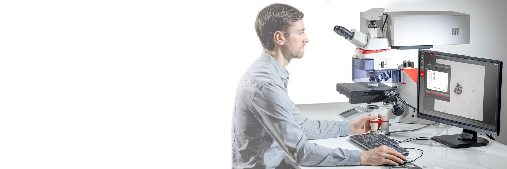
顕微鏡用画像解析ソフトウェア
顕微鏡用画像解析ソフトウェア
ライカ マイクロシステムズの画像解析ソフトウエアは、顕微鏡、デジタルカメラ、アクセサリーのトータルシステムとして制御可能です。
おすすめ製品 Show subnavigation
顕微鏡用画像解析ソフトウェア
ライカの顕微鏡アプリにより、イメージングや記録が簡単になります。 ライカのソフトウェアは、クリニカル、工業、教育用など、広くお使いいただいています。
ライカサイエンスラボ Show subnavigation
顕微鏡イメージングソフトウェア に関する最新の記事を読む
ライカ マイクロシステムズのサイエンスラボでは、 顕微鏡に関する専門記事やお客様事例、基本情報を提供しています。 日常業務や研究で、ビギナーから経験豊富な方まで幅広くサポートします。
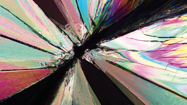
The Polarization Microscopy Principle
Polarization microscopy is routinely used in the material and earth sciences to identify materials and minerals on the basis of their characteristic refractive properties and colors. In biology, polarization microscopes are commonly used for identification of birefringent structures, like crystals, or for imaging of cellulose in the walls of plant cells and starch grains. This article gives an overview of the basic principles of polarization microscopy.

Super-Resolution Microscopy Image Gallery
Due to the diffraction limit of light, traditional confocal microscopy cannot resolve structures below ~240 nm. Super-resolution microscopy techniques, such as STED, PALM or STORM or some deconvolution processing methods, are used when enhanced resolution is needed to study structures and molecular events below the diffraction limit scale.

Extended Live-cell Imaging at Nanoscale Resolution
Extended live-cell imaging with TauSTED Xtend. Combined spatial and lifetime information allow super-resolution microscopy at extremely low light dose.
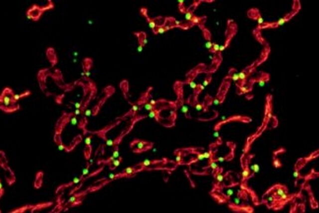
Studying Virus Replication with Fluorescence Microscopy
The results from research on SARS-CoV-2 virus replication kinetics, adaption capabilities, and cytopathology in Vero E6 cells, done with the help of fluorescence microscopy, are described in this article.
Epi-Illumination Fluorescence and Reflection-Contrast Microscopy
This article discusses the development of epi-illumination and reflection contrast for fluorescence microscopy concerning life-science applications. Much was done by the Ploem research group collaborating with the company Leitz.
Going Beyond Deconvolution
Widefield fluorescence microscopy is often used to visualize structures in life science specimens and obtain useful information. With the use of fluorescent proteins or dyes, discrete specimen components are marked in a highly specific manner. To fully understand a structure, visualizing it in 3 dimensions can be necessary, but certain challenges are faced when doing so with microscopy.
AI Microscopy Image Analysis – An Introduction
Artificial intelligence-guided microscopy image analysis and visualization is a powerful tool for data-driven scientific discovery. AI can help researchers tackle challenging imaging applications, allowing them to extract more information from their images.
The Potential of Coherent Raman Scattering Microscopy at a Glance
Coherent Raman scattering microscopy (CRS) is a powerful approach for label-free, chemically specific imaging. It is based on the characteristic intrinsic vibrational contrast of molecules in the sample.
CRS provides high-resolution (sub-cellular level) and dynamic (up to video rate) information on the biochemical composition and metabolic processes in cells, tissues, and intact model organisms. It also enables imaging of small molecules without perturbing their function. This information is highly synergistic with the molecular contrast provided by fluorescence microscopy. Unsurprisingly, CRS is finding a growing number of applications in fields like neurodegenerative disease, cancer, 3D biology, stem cell and developmental biology, and pharmacology.
Simplifying Complex Fluorescence Multiwell Plate Assays
Apoptosis, or programmed cell death, occurs during organism embryo development to eliminate unwanted cells and during healing in adults to rid the body of damaged cells and help prevent cancer. Caspase assay experiments with multiwell plates allow researchers to study the early phases of apoptosis. In this article, we show how Mica can be used in this key application with fluorescence multiwell plate assays to provide 100% spatiotemporal correlation of data and minimize crosstalk .
Efficient Long-term Time-lapse Microscopy
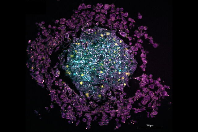
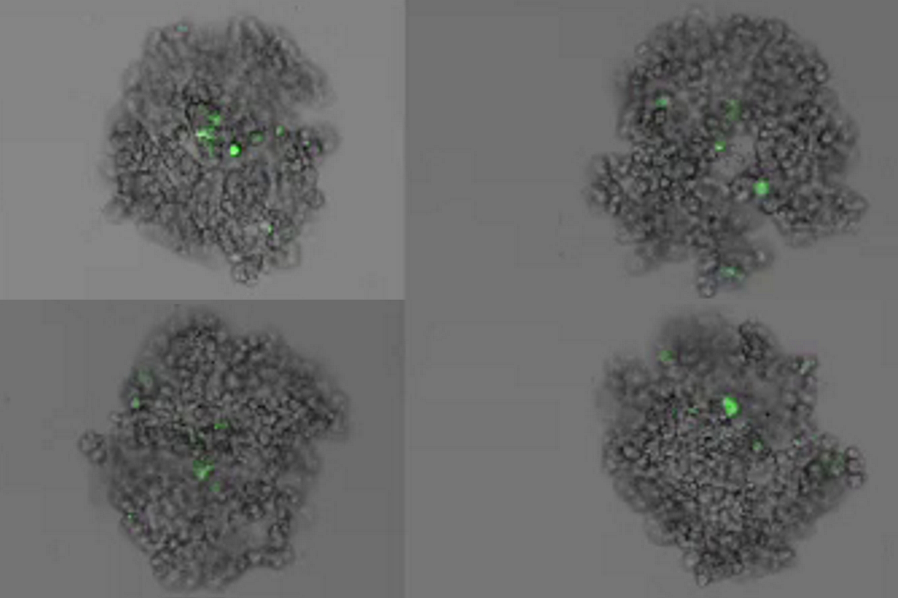
When doing time-lapse microscopy experiments with spheroids, there are certain challenges which can arise. As the experiments can last for several days, prolonged sample survival must be achieved which requires that near physiological conditions are ensured. The long-term time-lapse study described in this article involves the use of Mica - the world’s first Microhub, to investigate the formation of spheroids from U343 and MDCK cells. Growing spheroids requires optimal conditions to ensure undisturbed cell cycles and proliferation.
Multicolor 4D Super Resolution Light Sheet Microscopy
The AI Microscopy Symposium offers a unique forum for discussing the latest AI-based technologies and tools in the field of microscopy and biomedical imaging. In this scientific presentation, Yuxuan Zhao demonstrates how 3D imaging of organelles within live cells can be improved using a progressive deep learning strategy combined with a dual-ring-modulated SPIM design.
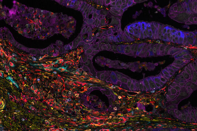
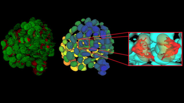
Hyperplex Cancer Tissue Analysis at Single Cell Level with Cell DIVE
The ability to study how lymphoma cell heterogeneity is influenced by the cells’ response to their microenvironment, especially at the mutational, transcriptomic, and protein levels. Protein expression studies offer the most relevant information about the nature of cellular interactions and protein expression levels. A hyperplexed workflow can be applied for studying multiple proteins from the same cancer tissue.
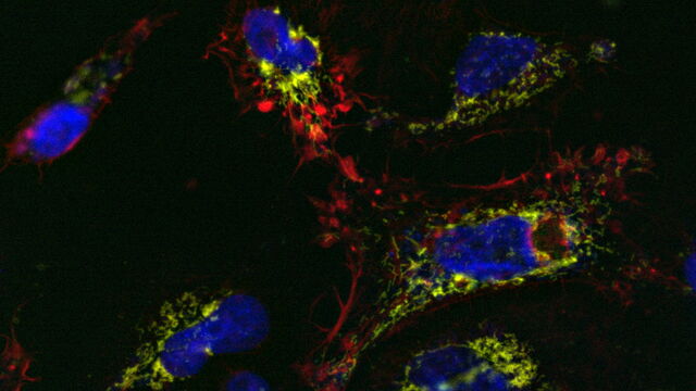
How to Prepare your Specimen for Immunofluorescence Microscopy
Immunofluorescence (IF) is a powerful method for visualizing intracellular processes, conditions and structures. IF preparations can be analyzed by various microscopy techniques (e.g. CLSM, Epifluorescence, TIRF, GSDIM), depending on the application or the researcher’s interest. Meanwhile, IF has become indispensable for a large number of research groups which have at least access to a simple fluorescence microscope.
Live-Cell Imaging Techniques
The understanding of complex and/or fast cellular dynamics is an important step for exploring biological processes. Therefore, today’s life science research is increasingly focused on dynamic processes like cell migration, morphological changes of cells, organs, or whole animals, and real-time physiological (e.g., changes of intracellular ion composition) events in living specimens. One approach to address these challenging demands is to employ certain optical methods that are collectively called live-cell imaging.
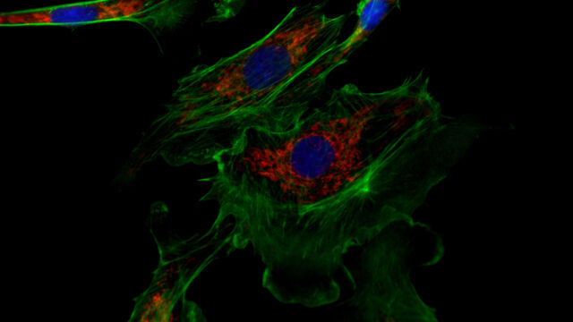
Fluorescent Dyes
A basic principle in fluorescence microscopy is the highly specific visualization of cellular components with the help of a fluorescent agent. This can be a fluorescent protein – for example GFP – genetically linked to the protein of interest. If cloning is impossible – for instance in histologic samples – techniques such as immunofluorescence staining are used to visualize the protein of interest.
The AI-Powered Pixel Classifier
Achieving reproducible results manually requires expertise and is tedious work. But now there is a way to overcome these challenges by speeding up this analysis to extract the real value of the image and gain insights. The artificial-intelligence-powered pixel classifier provides reproducible segmentation results fast and overcomes human action. It delivers more robust results compared to classical function-based automation.
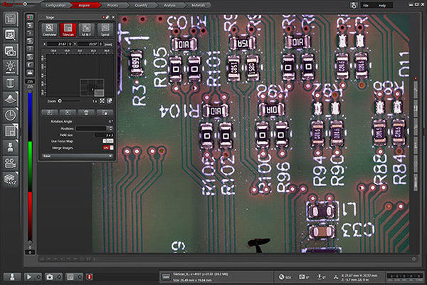
コア機能
- 顕微鏡とデジタルカメラの構成と制御を完全に統合。
- 注釈ツールにより、画像データと較正データを画像に追加できます。
- カメラの露出調整で、イメージングの条件を最適化できます。
- 素早く簡単に確認できる、取得したイメージのサムネイルギャラリー。
- ライカの顕微鏡とカメラのデータを使用して、画像は自動的に較正されます。スケールバーは画像のサイズを示します。
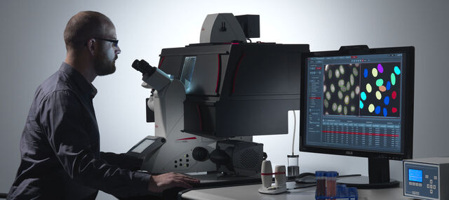
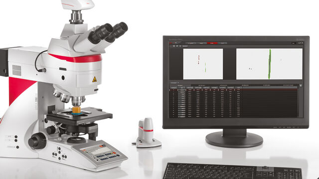
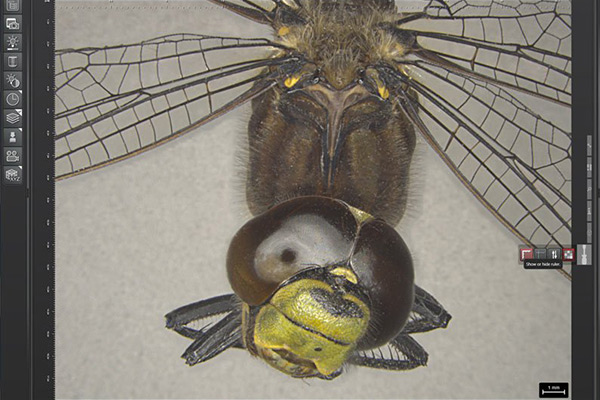
LAS X Industry
- フルスクリーンでサンプルを観察
- 解析時の測定を効率的かつ柔軟に
- イメージングをX、Y、Z軸で強化
- 信頼、再現可能な結果を得る
- ニーズに合わせてレポートテンプレートをカスタマイズ
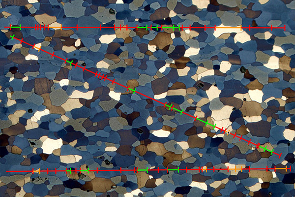
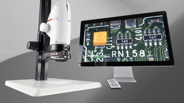
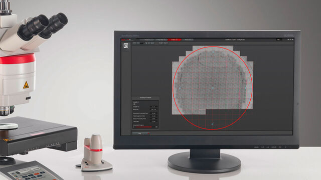
LAS Xマテリアルモジュール
- 鋼やその他マテリアルの構造分析を迅速に実施できます。
- 金属組織学(鉄鋼/鋳物製品)向けの画像解析を可能にします。
- さまざまな工業規格に従って、金属や合金の微細組織を手動、自動分析します。
- 人間工学を大幅に改善し、長時間の観察でも高い精度で分析を実施できます。
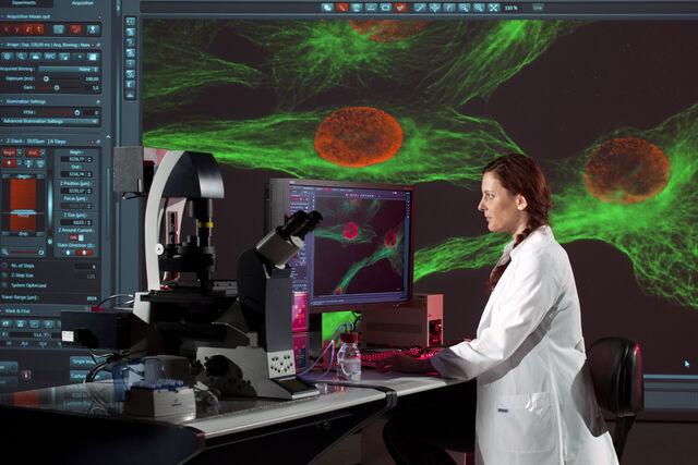
LAS Xライフサイエンス
- ライカ マイクロシステムズの共焦点顕微鏡、広視野顕微鏡、実体顕微鏡、超解像顕微鏡、ライトシート顕微鏡を統合。
- 細胞のGPS「LAS X Navigator」が高速で全体像を捉え、瞬時に重要な細部を特定
- イメージングタスクが直感的になり、研究に集中できる
