Kontaktieren Sie uns

David R. Barbero , PhD

David R. Barbero

Dr. David R. Barbero ist Ingenieur und Physiker und verfügt über mehr als 15 Jahre Erfahrung mit optoelektronischen Materialien, Geräten und Displays im Nanobereich. Seine Arbeiten wurden in mehreren führenden Fachzeitschriften für Materialwissenschaft und Physik veröffentlicht und in über 300 wissenschaftlichen Zeitungen und Internet-Websites vorgestellt. Er verfügt über langjährige Erfahrung in der Nanomusterung und Nanostrukturierung von Polymeren und Verbundwerkstoffen (optische Lithografie, Nanoimprinting, kolloidale Nanoröhren und Polymerselbstorganisation). David promovierte in Physik am Cavendish Laboratory der Universität Cambridge (Vereinigtes Königreich), wo er als Marie-Curie-Stipendiat und European Cambridge Trust Scholar tätig war. 2009 wurde er mit dem Abdus-Salam-Zweitplatzierten-Preis der Universität Cambridge in Physik ausgezeichnet.

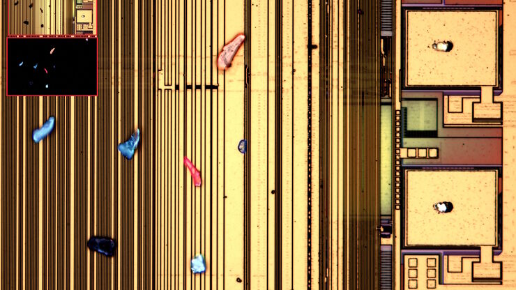
Particulate contamination in between moving metal plates.

Key Factors for Efficient Cleanliness Analysis

An overview of the key factors necessary for technical cleanliness and efficient cleanliness analysis concerning automotive and electronics manufacturing and production is provided in this article.
Images of the same area of a processed wafer taken with standard (left) and oblique (right) brightfield illumination using a Leica compound microscope. The defect on the wafer surface is clearly more visible with oblique illumination.

Rapid Semiconductor Inspection with Microscope Contrast Methods

Semiconductor inspection for QC of materials like wafers can be challenging. Microscope solutions that offer several contrast methods enable fast and reliable defect detection and efficient workflows.
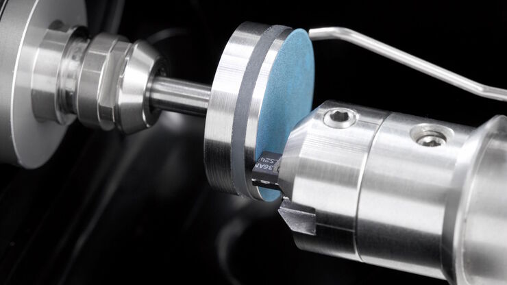
Preparation of an IC-chip cross section: grinding and polishing of the chip cross section.

Cross-section Analysis for Electronics Manufacturing

This article describes cross-section analysis for electronics concerning quality control and failure analysis of printed circuit boards (PCBs) and assemblies (PCBAs), integrated circuits (ICs), etc.
Chicken-embryo cross section at the level of the midbrain showing neural crest cells and cadherin-6B molecules. Raw widefield data (top) and THUNDER image (bottom).

The Neural Crest (NC)

This article discusses how the study of neural crest (NC) development in chicken embryos is aided with haze-free imaging using a THUNDER Imager 3D Assay. Proper specification, migration, and…
Raw widefield and THUNDER image of transversal mouse adult fiber lens section. Courtesy N. Houssin, Plagemen lab, Ohio State University, Columbus, USA.

Studying Ocular Birth Defects

This article discusses how lens formation and ocular birth defects can be studied with sharp widefield microscopy images which are acquired rapidly. The mouse ocular lens is used as a model to study…
THUNDER image of early prophase Drosophila germarium.

Healthy Oocyte Development

This article discusses how the study of mechanisms involved in the generation of healthy oocytes in Drosophila fruit flies can be helped with sharp, haze-free images acquired with a THUNDER Imager…
The various solutions from Leica Microsystems for cleanliness analysis.

Factors to Consider for a Cleanliness Analysis Solution

Choosing the right cleanliness analysis solution is important for optimal quality control. This article discusses the important factors that should be taken into account to find the solution that best…
Chicken cochlea tissue imaged with a THUNDER Imager using large volume computational clearing. Courtesy of Dr. Amanda Janesick, California, USA.

Development and Recovery of the Inner Ear

This article discusses how thick cochlear tissues of chicken embryos can be sharply imaged for studying inner-ear hair cell regeneration. Sensory hair cells perform important functions for hearing and…
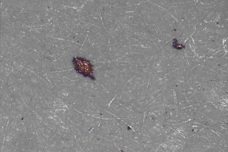
[Translate to German:] Particles and fibers on a filter which will be counted and analyzed for cleanliness

Effiziente Partikelzählung und -analyse

Dieser Bericht befasst sich mit der Partikelzählung und -analyse unter Verwendung der optischen Mikroskopie bei der technischen Sauberkeitsanalyse von Teilen und Komponenten. Die Partikelzählung und…
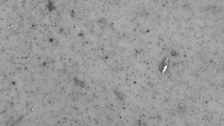
[Translate to German:] Particles which could be found during cleanliness analysis of parts and components.

Technische Sauberkeit von Automobilkomponenten und -teilen

In diesem Artikel werden die ISO-Norm 16232 und die VDA 19-Richtlinien erläutert und die Verfahren zur Partikelanalyse kurz zusammengefasst. Diese liefern wichtige Kriterien für die Sauberkeit von…
Macroscopic extended depth of field images of C. elegans

Autophagy and Age-Related Pathologies

High-contrast imaging of the model organism C. elegans, a nematode, with the THUNDER Imager Model Organism using Small Volume Computational Clearing (SVCC) helps to better understand how autophagy and…
Electronic component

Top Challenges for Visual Inspection

This article discusses the challenges encountered when performing visual inspection and rework using a microscope. Using the right type of microscope and optical setup is paramount in order to…
[Translate to German:]  Routine inspection microscope Ivesta 3

How to select the right solution for visual inspection

This article helps users with the decision-making process when selecting a microscope as a solution for routine visual inspection. Important factors that should be considered are described.

Role of Mucins and Glycosylation in Dry Eye Disease

This article shows how fast, high-contrast, and sharp imaging of stratified human corneal epithelial cells with THUNDER imaging technology for dry eye disease (DED) research allows membrane ridges to…
Images of C2C12 cells which were stained with lamin B (magenta), Hoechst (blue), and yH2AX (yellow). A) raw widefield fluorescence image and B) THUNDER image after LVCC. The yellow foci indicate damaged DNA.

Skeletal Muscle Adaptation and Fibrotic Diseases

The mechanisms of how skeletal muscle adapts to fibrotic pathologies can be investigated more efficiently with fast, high-contrast imaging of C2C12 mouse myoblast cells which is described in this…

Introduction to 21 CFR Part 11 and Related Regulations

This article provides an overview of regulations and guidelines for electronic records (data entry, storage, signatures, and approvals) used in the USA (21 CFR Part 11), EU (GMP Annex 11), and China…
Images of a brain organoid derived from iPSCs acquired with a THUNDER Imager 3D Cell Culture. The cells were infected with the pAAV-hSyn-EGFP and pLX-hGFAP-mCherry virus. The image is the 36th plane cropped out of a 53 plane Z-stack volume. Shown are both the A) raw widefield image and B) the same image after Large Volume Computation Clearing (LVCC). Neurons are labeled in green and astrocytes in red.

“Brains-In-A-Dish” from Induced Pluripotent Stem Cells (iPSCs)

This article discusses the benefits of using the THUNDER technology for imaging inside 3D human cortical brain organoids. These organoids are derived from human induced pluripotent stem cells (iPSCs)…

Why is Manual Visual Inspection of Medical Devices so Challenging?

This article discusses how manual visual inspection, which is prevalent in the medical device industry, can lead to inconsistent results. It also addresses the challenges quality managers and…
Scroll to top