The microstructure of polymers controls their chemical reactivity as well as their mechanical and transport properties. However, quantitative characterization of the microstructure of soft polymers is not often possible or practical with the few analytical methods available to study it at the sub-micrometer scale. Freeze-fracturing is one such method, but often the surfaces of samples prepared with the process are too rough, leaving only a very small area appropriate for quantitative SEM investigation.
The results demonstrate that cryo-BIB-SEM enables microstructure characterization of soft polymers with high resolution over large planar areas of damage-free sample cross sections. Only minor artifacts from the cryo-BIB-SEM sample preparation process were seen at the scale of observation, none of which appear to be a consequence of freezing.
Introduction
Precise characterization of a polymer’s microstructure is important, because it influences the chemical reactivity and mechanical and transport properties of the polymer and, thus, governs durability and robustness to weathering, heat, cold, and light exposure.
Moreover, this fact applies to any type of polymer, whether soft, hard, natural, or synthetic.
Cryo-BIB-SEM was developed to enable high resolution imaging and chemical analysis of micropores over relatively large, cryogenically stabilized samples with damage-free areas. In a previous report, cryo-BIB-SEM was used to study the microstructure of lithium-ion battery electrodes during drying [1]. Here, cryo-BIB-SEM is applied to characterize natural polymers. Examples of natural polymers are wood and the skin of fruits and vegetables.
As mentioned above, an accurate determination of the soft polymer microstructure with sub-micrometer resolution is necessary. Unfortunately, few methods can deliver such a high resolution for such soft samples. Furthermore, quantitatively characterizing the microstructure of freeze-fractured soft polymer samples is often difficult, because the surfaces roduced are too rough making analysis with SEM or energy dispersive spectroscopy (EDS) challenging as only small areas can be accurately investigated. Additionally, for organic materials which can swell and shrink, sample preparation for and measurement with SEM, especially materials with a high moisture content, are difficult to perform with the sample in its native state. Alternative methods, such as micro-computed tomography (μCT) and cryogenic focused ion beam milling with SEM (cryo-FIB-SEM), are often too low in resolution or the results obtained are not representative.
This application note describes the use of cryo-BIB-SEM to characterize at high resolution (sub-micrometer level) the microstructure of soft, delicate natural polymers (wood and tomato skin).
Materials and Methods Cryo-BIB-SEM
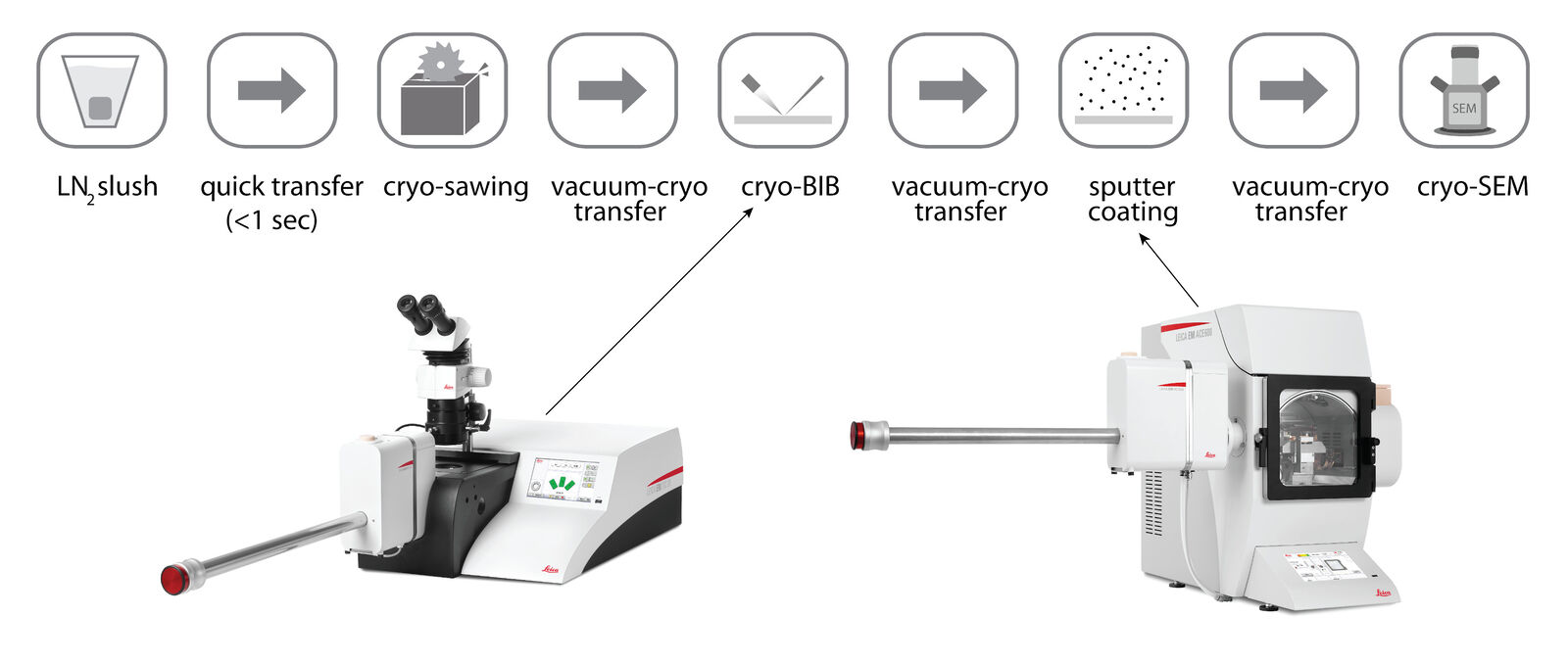
Cryo-BIB enables the preparation of cryogenically stabilized cross sections with a large planar area (as much as 4 mm²). The sample preparation uses a rapid cooling (quenching) step which allows a clean cut of the polymer with much less risk of damage, i.e., fracture, deformation, etc. The sample is quenched in a liquid nitrogen (LN2) slush and quickly transferred onto a cryogenic cooling stage where it is maintained at LN2 temperatures (Fig. 1a) [2]. A diamond blade saw is used to cut the cryogenically cooled sample a few tens of nm above a titanium (Ti) mask which shields the sample during the subsequent sputter coating process. Milling with an argon (Ar) ion beam using cryo-BIB produces a large, flat cross section.
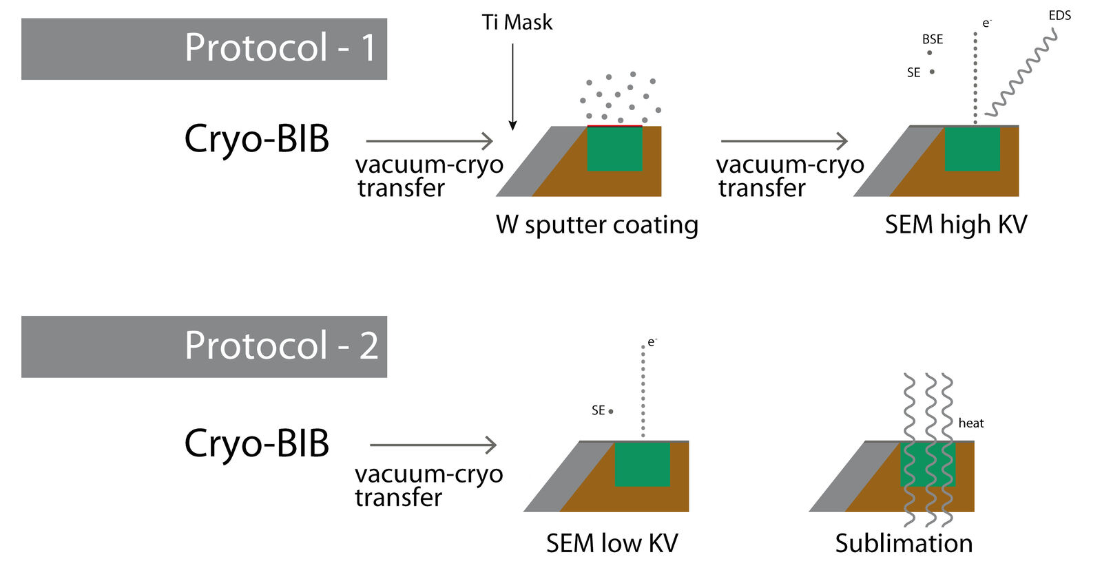
Next, two preparation and imaging protocols can be used, depending on the type of information needed, whether composition data from
EDS or high-resolution imaging of fine structures with SEM. In the first protocol (Fig. 1b), the sample is sputter coated to facilitate EDS/SEM analysis and provide information about the texture and composition of the phases. In the second protocol, the sample is not coated to allow for sublimation of any water which has infiltrated its porous surface. This way, any part of the sample microstructure, which was previously hidden by the water, can now be visualized.
Cryogenic broad ion beam (cryo-BIB) milling of wood and tomato skin (natural polymers) samples was done with the EM TIC 3X ion beam milling system from Leica Microsystems. Before the BIB-milled samples were placed in the SEM (Supra 55, Zeiss), they were sputter coated with a thin layer of tungsten (W) using the EM ACE600 sputter, carbon, and e-beam coater, also from Leica Microsystems. The W layer avoids charging of the non-conductive samples when irradiated by the electron beam.
Results
The main purpose of analyzing soft natural polymers was to validate the ability of the cryo-BIB-SEM method to preserve fine and delicate structures during sample preparation and analysis. In particular, one goal was to determine the quality of the cross section produced by the cryo-BIB-SEM sample preparation process, i.e., if artifacts or damage are introduced on the surface of the sample cross section.
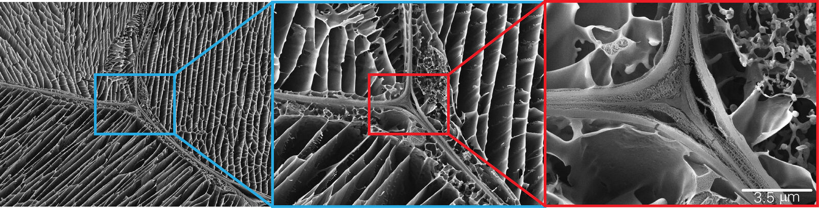
Tomato Skin
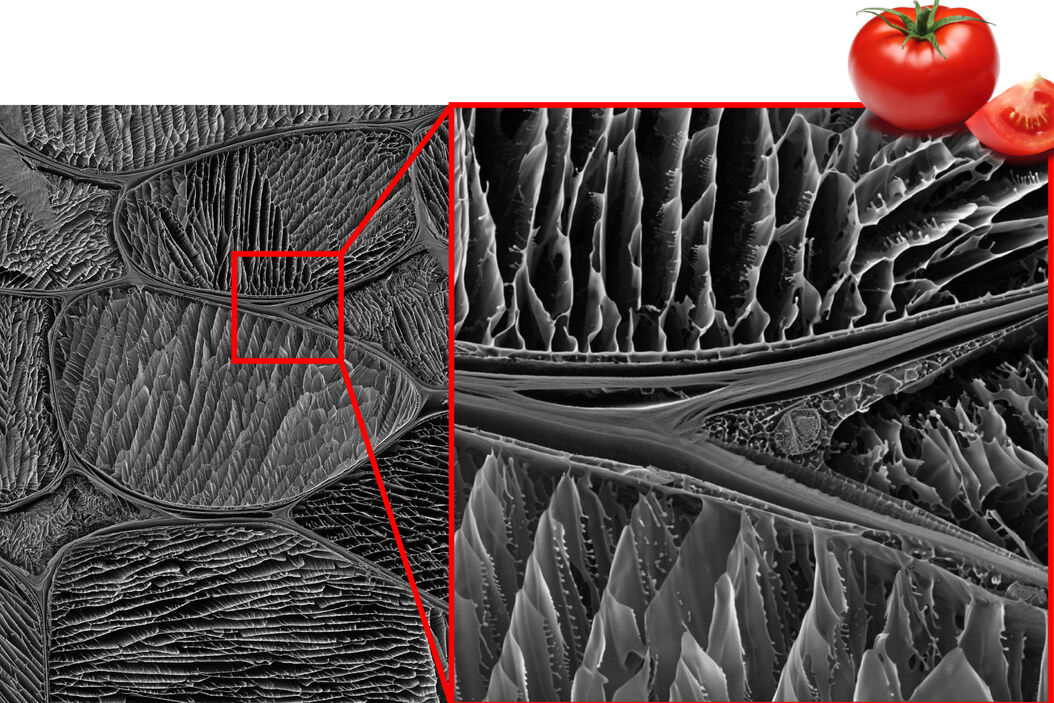
A small piece of skin was cut from a fresh tomato (Fig. 2a) which was then prepared according to the workflow in figure 1 and analyzed according to protocol 2. Tomato fruit contains a lot of water and has a very complex microstructure, therefore, it is an ideal material for assessing the potential for fractures due to ice crystals forming during the sample preparation.
Figure 2b shows a cross section of the tomato skin, where a small area of the Ti mask can be seen on top of the cross section which has collected some “sputter dust”. The fine structure of the tomato cells is clearly visible and the cell shape is very similar to what has been previously reported in the literature from optical microscopy observations [3]. At higher resolution, the cryo-BIB-SEM method enabled even delicate microstructures to be analyzed in fine detail (Fig 2c).
No evidence of damage resulting from the cryogenic-cooling and cutting process could be found, which highlights the ability of the cryo-BIB-SEM method to produce pristine, defect-free samples for analysis of the microstructure of a soft, delicate natural polymer.
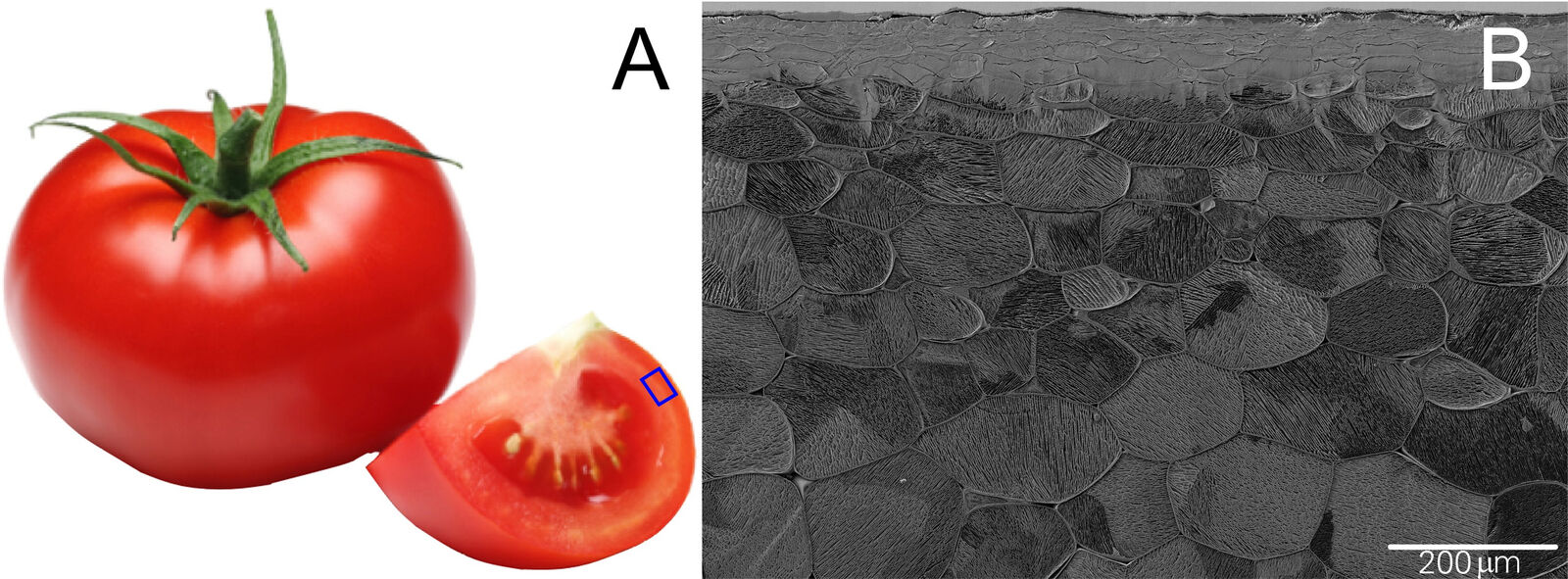
2b: Cryo-BIB-SEM image showing a large area of a tomato skin cross-section sample.
Cryo-BIB-SEM imaging can reveal the cells and microscale morphology of pine wood (Pinus sylvestris). Broad ion beam milling produces a cell wall surface without artifacts caused by preparation techniques like microtome cutting. EDS provides the composition of the different wood phases, for example carbon (C) present in the cellulose and oxygen (O) from the large amount of water in the cells [4].
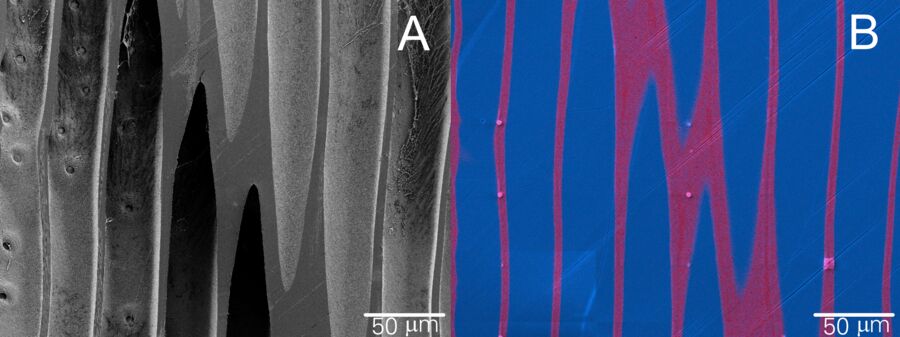
3b: EDS image of cryo-BIB prepared pine wood (same area as seen in 3a). The carbon (C, red) signal comes from the cellulose and oxygen (O, blue) from water in the wood cells.
Summary and Conclusion
In summary, we have shown that cryo-BIB-SEM is a powerful characterization method which can be used to investigate cross sections of delicate, soft natural polymers (tomato skin and wood). The fine details of the microstructure of undamaged tomato skin and wood cells could be imaged with high resolution. Moreover, no evidence of fracture or sample damage due to the cryo-BIB-SEM sample preparation process was observed.
References
- S. Jaiser, J. Kumberg, J. Klaver, J.L. Urai, W. Schabel, J. Schmatz, P. Scharfer, Microstructure formation of lithium-ion battery electrodes during drying - An ex-situ study using cryogenic broad ion beam slope-cutting and scanning electron microscopy (Cryo-BIB-SEM), J. Power Sources (2017) vol. 345, pp. 97-107, DOI: 10.1016/j. jpowsour.2017.01.117
- J. Schmatz, J. Klaver, M. Jiang, J.L. Urai, Nanoscale Morphology of Brine/Oil/Mineral Contacts in Connected Pores of Carbonate Reservoirs: Insights on Wettability From Cryo-BIB-SEM, SPE Journal (2017) vol. 22, iss. 05, DOI: 10.2118/180049-PA.
- R. Metzner, H.U. Schneider, U. Breuer, W.H. Schroeder, Imaging Nutrient Distributions in Plant Tissue Using, Time-of-Flight Secondary Ion Mass Spectrometry and Scanning Electron Microscopy, Plant Physiology (2008) vol. 147, pp. 1774–1787, DOI: 10.1104/ pp.107.109215.
- M. Nopens, J. Schmatz, Saturated pine wood sample, Pinus sylvestris, 2017, MaP Microstructures and Pores.
Related Articles
-
 labeled with membrane-permeable calcein, high-pressure frozen in salt water using EM ICE.")
High-Pressure Freezing for Organoids: Cryo CLEM & FIB Lift Out
Master cryo EM workflow steps for challenging 3D samples: when to choose HPF vs. plunge freezing,…
Jan 22, 2026Read article -

Integrated Serial Sectioning and Cryo-EM Workflows for 3D Biological Imaging
This on-demand webinar explores how integrated tools can support electron microscopy workflows from…
Jul 22, 2025Read article -

Revealing Sodium Battery Degradation via Cryo-EM and CryoFIB
Explore how cryogenic electron microscopy and focused ion beam techniques uncover the intrinsic…
Jul 22, 2025Read article
