David R. Barbero , PhD

Dr. David R. Barbero is an engineer and physicist who has over 15 years of experience in nanoscale opto-electronic materials, devices and displays. His work has been published in several top journals in Materials Science and Physics and highlighted in over 300 scientific newspapers and internet websites. He has a long experience in nano-patterning and nano-structuring of polymers and composites (optical lithography, nano-imprinting, colloidal, nanotubes, and polymer self-assembly). David obtained his PhD in Physics at the Cavendish Laboratory, University of Cambridge, UK, where he was a Marie-Curie Fellow, a European Cambridge Trust Scholar, and he was awarded the Abdus Salam runner-up prize in Physics from Cambridge University in 2009.

How to Select the Right Measurement Microscope
With a measurement microscope, users can measure the size and dimensions of sample features in both 2D and 3D, something crucial for inspection, QC, failure analysis, and R&D. However, choosing the…

Microscope Calibration for Measurements: Why and How You Should Do It
Microscope calibration ensures accurate and consistent measurements for inspection, quality control (QC), failure analysis, and research and development (R&D). Calibration steps are described in this…

Visualizing Photoresist Residue and Organic Contamination on Wafers
As the scale of integrated circuits (ICs) on semiconductors passes below 10 nm, efficient detection of organic contamination, like photoresist residue, and defects during wafer inspection is becoming…

Rapidly Visualizing Magnetic Domains in Steel with Kerr Microscopy
The rotation of polarized light after interaction with magnetic domains in a material, known as the Kerr effect, enables the investigation of magnetized samples with Kerr microscopy. It allows rapid…
 at the edge of a battery electrode acquired with a DVM6 digital microscope.")
Burr Detection During Battery Manufacturing
See how optical microscopy can be used for burr detection on battery electrodes and determination of damage potential to achieve rapid and reliable quality control during battery manufacturing.

Introduction to 21 CFR Part 11 for Electronic Records of Cell Culture
This article provides an introduction to the recommendations of 21 CFR Part 11 from the FDA, specifically focusing on the audit trail and user management in the context of cell-culture laboratories.…

Battery Particle Detection During the Production Process
How battery particle detection and analysis is enhanced with optical microscopy and laser spectroscopy for rapid, reliable, and cost-effective QC during battery production is explained in this…

Key Factors for Efficient Cleanliness Analysis
An overview of the key factors necessary for technical cleanliness and efficient cleanliness analysis concerning automotive and electronics manufacturing and production is provided in this article.
 and oblique (right) brightfield illumination using a Leica compound microscope. The defect on the wafer surface is clearly more visible with oblique illumination.")
Rapid Semiconductor Inspection with Microscope Contrast Methods
Semiconductor inspection during the production of patterned wafers and ICs (integrated circuits) is important for identifying and minimizing defects. To increase the efficiency of quality control in…

Quality Control via Cross Sections of PCBs, PCBAs, ICs, and Batteries
Why cross sections of printed circuit boards (PCBs) and assemblies (PCBAs), integrated circuits (ICs), and battery components are useful for quality control (QC), failure analysis (FA), and research…

Top Challenges for Visual Inspection
This article discusses the challenges encountered when performing visual inspection and rework using a microscope. Using the right type of microscope and optical setup is paramount in order to…

3 Factors Determine the Damage Potential of Particles
This article discusses the 3 factors for determining the potential of a particle to cause damage to parts and components in the automotive and electronic industry. These factors include the…

Factors to Consider for a Cleanliness Analysis Solution
Choosing the right cleanliness analysis solution is important for optimal quality control. This article discusses the important factors that should be taken into account to find the solution that best…

Efficient Particle Counting and Analysis
This report discusses particle counting and analysis using optical microscopy for cleanliness of parts and components. Particle counting and analysis is a critical part of quality assurance in the…

Cleanliness of Automotive Components and Parts
This article discusses the ISO 16232 standard and VDA 19 guidelines and briefly summarizes the particle analysis methods. They give important criteria for the cleanliness of automotive parts and…

How to Select the Right Solution for Visual Inspection
This article helps users with the decision-making process when selecting a microscope as a solution for routine visual inspection. Important factors that should be considered are described.

Keeping Particulate Contamination Under Control in Pharmaceutical Products
This article describes how a 2-methods-in-1 solution combining optical microscopy and laser induced breakdown spectroscopy (LIBS) can be utilized for identification of particulate contaminants in the…

How does an Automated Rating Solution for Steel Inclusions Work?
The rating of non-metallic inclusions (NMIs) to determine steel quality is critical for many industrial applications. For an efficient and cost-effective steel quality evaluation, an automated NMI…

Challenges Faced When Manually Rating Non-Metallic Inclusions (NMIs) to Determine Steel Quality
Rapid, accurate, and reliable rating of non-metallic inclusions (NMIs) is instrumental for the determination of steel quality. This article describes the challenges that arise from manual NMI rating,…

Top Issues Related to Standards for Rating Non-Metallic Inclusions in Steel
Supplying components and products made of steel to users worldwide can require that a single batch be compliant with multiple steel quality standards. This user demand creates significant challenges…
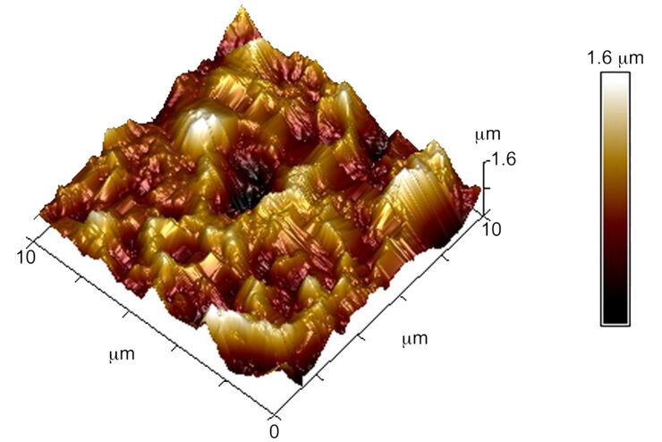
Brief Introduction to Surface Metrology
This report briefly discusses several important metrology techniques and standard definitions commonly used to assess the topography of surfaces, also known as surface texture or surface finish. With…
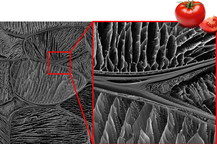
Studying the Microstructure of Natural Polymers in Fine Detail
The potential of cryogenic broad ion beam milling used in combination with scanning electron microscopy (cryo-BIB-SEM) for imaging and analyzing the microstructure of cryogenically stabilized soft…
