Leica Microsystems

Leica Microsystems

Leica Microsystems ist ein weltweit führender Hersteller von Mikroskopen und wissenschaftlichen Instrumenten. Im 19. Jahrhundert als Familienunternehmen gegründet, war die Geschichte des Unternehmens auf dem Weg zum Weltkonzern von beispielloser Innovation geprägt.

Die traditionell enge Zusammenarbeit mit der Wissenschaft ist der Schlüssel zur Innovationstradition von Leica Microsystems, die auf die Ideen der Anwender zurückgreift und auf deren Bedürfnisse zugeschnittene Lösungen schafft. Auf globaler Ebene ist Leica Microsystems in drei Divisionen gegliedert, die alle zu den führenden Anbietern in ihrem jeweiligen Bereich gehören: Life Science, Industrie und Medizin.

Das Unternehmen ist in über 100 Ländern mit 6 Produktionsstandorten in 5 Ländern, Vertriebs- und Serviceorganisationen in 20 Ländern und einem internationalen Händlernetz vertreten. Der Hauptsitz des Unternehmens liegt in Wetzlar, Deutschland.
https://www.leica-microsystems.com/de

Jellyfish Aequorea Victoria

Fluorescent Proteins - From the Beginnings to the Nobel Prize

Fluorescent proteins are the fundament of recent fluorescence microscopy and its modern applications. Their discovery and consequent development was one of the most exciting innovations for life…
Snapshot from a time lapse of a calcium imaging experiment using the ratiometric calcium indicator Fura-2.

Ratiometric Imaging

Many fundamental functions of a cell strongly depend on delicate, but nevertheless dynamic balances of ions (e.g. calcium, magnesium), voltage potentials and pH between the cell’s cytosol and the…

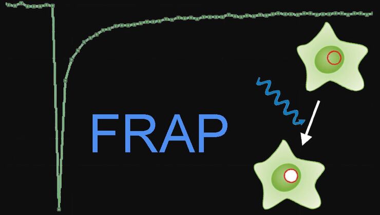
Fluorescence Recovery after Photobleaching (FRAP) and its Offspring

FRAP (Fluorescence recovery after photobleaching) can be used to study cellular protein dynamics: For visualization the protein of interest is fused to a fluorescent protein or a fluorescent dye. A…

Förster Resonance Energy Transfer (FRET)

The Förster Resonance Energy Transfer (FRET) phenomenon offers techniques that allow studies of interactions in dimensions below the optical resolution limit. FRET describes the transfer of the energy…

An Introduction to CARS Microscopy

CARS overcomes the drawbacks of conventional staining methods by the intrinsic characteristics of the method. CARS does not require labeling because it is highly specific to molecular compounds which…
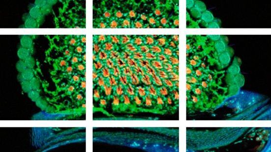
Scheme of a 2D mosaic scan. Drosophila melanogaster (eye section)

Mosaic Images

Confocal laser scanning microscopes are widely used to create highly resolved 3D images of cells, subcellular structures and even single molecules. Still, an increasing number of scientists are…
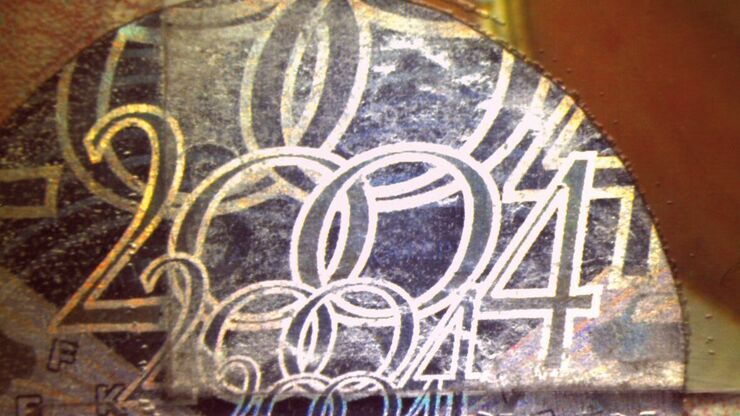
An ID card which has been tampered with by counterfeiters who inserted a hologram.

Is that Document Genuine or Fake? How do They Identify Fake Documents?

This article shows how forensic experts use microscopy for analysis to identify counterfeit, fake documents, such as ID cards, passports, visas, certificates, etc. Then they know if it is genuine or…

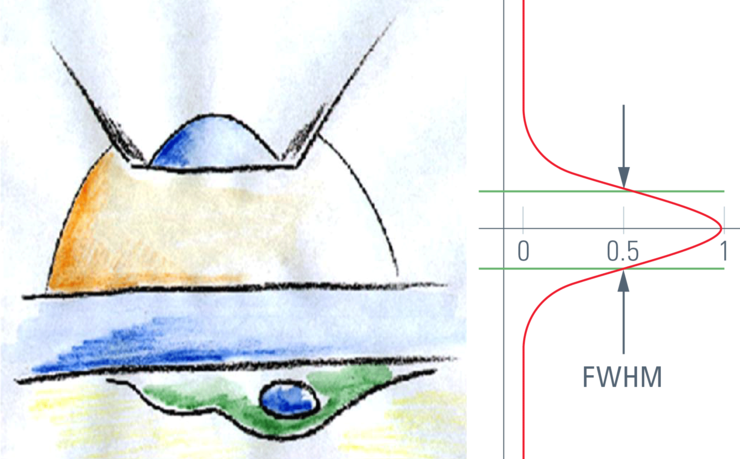
Confocal Optical Section Thickness

Confocal microscopes are employed to optically slice comparably thick samples.
Der Modulationskontrast visualisiert transparente kontrastarme Proben

Integrated Modulation Contrast (IMC)

Hoffman modulation contrast has established itself as a standard for the observation of unstained, low-contrast biological specimens. The integration of the modulator in the beam path of themodern…
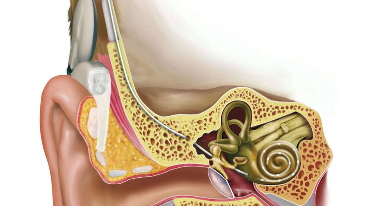
Cochlea implant. Illustration: © MED-EL.

Cochlea Implants for Deaf and Severely Hard of Hearing

The cochlea implant (CI) replaces the function of the outer ear, the middle ear and the ­cochlea of the inner ear. Basically, it consists of an external unit comprising a microphone, speech processor,…

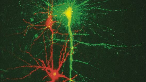
New Standard in Electrophysiology and Deep Tissue Imaging

The function of nerve and muscle cells relies on ionic currents flowing through ion channels. These ion channels play a major role in cell physiology. One way to investigate ion channels is to use…
A portion of an early binocular microscope developed by John Leonhard Riddel in the early 1850s.

The History of Stereo Microscopy

This article gives an overview on the history of stereo microscopes. The development and evolution from handcrafted instruments (late 16th to mid-18th century) to mass produced ones the last 150…
Scroll to top