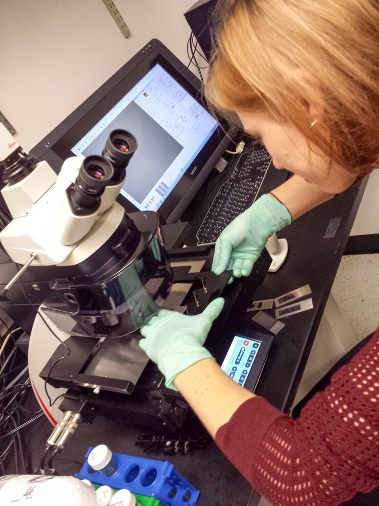
Leica LMD6 & LMD7
正立顕微鏡
光学顕微鏡
製品紹介
Home
Leica Microsystems
Leica LMD6 & LMD7 レーザーマイクロダイセクション
ダイセクションで完璧なカット
最新の記事を読む

Spatial Proteomics Workflow in Blood Cancer (MPNs)
Megakaryocytes play a central role in the biology of myeloproliferative neoplasms (MPNs), yet their in vivo proteomic characterization remains a major challenge due to low abundance and disrupted…
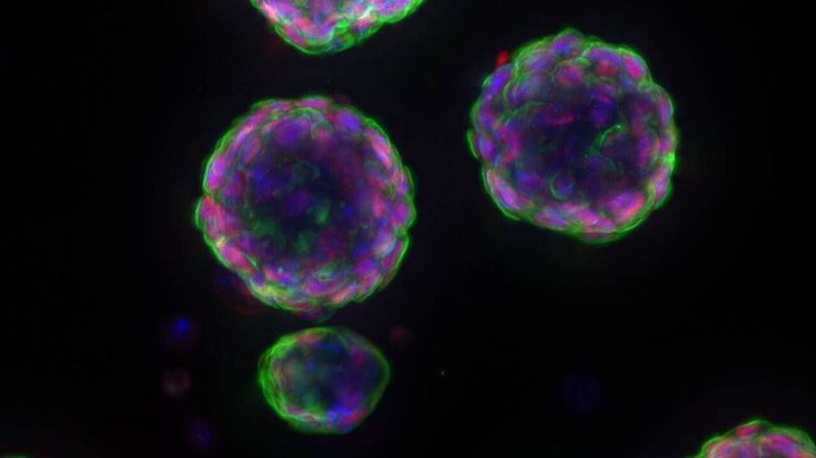
 and astrocytes (green) in a cortical spheroid derived from human induced pluripotent stem cells.")
Guide to Live-Cell Imaging
For a wide range of applications in various research fields of life science, live-cell imaging is an indispensable tool for visualizing cells in a state as close to in vivo, i.e. living and active, as…

Factors to Consider When Selecting a Research Microscope
An optical microscope is often one of the central devices in a life-science research lab. It can be used for various applications which shed light on many scientific questions. Thereby the…

AI meets Deep Visual Proteomics (DVP) to Advance Disease Research
In this webinar, Dr. Andreas Mund will introduce a cutting-edge platform that merges Deep Visual Proteomics (DVP) with AI-powered pathology models, enabling high-resolution mapping of key regions in…
 segmentation used in conjunction with LMD to increase discovery throughput.")
Biomarker Discovery with Laser Microdissection
Explore the potential of spatial proteomics workflows, such as Deep Visual Proteomics (DVP), to decipher pathology mechanisms and uncover druggable targets.
Altered protein expression, abundance, or…
 at Leica Microsystems.")
How a Breakthrough in Spatial Proteomics Saved Lives
Toxic epidermal necrolysis (TEN) is a rare but devastating reaction to common medications like antibiotics or gout treatments. It begins innocuously, often as a rash, but can escalate rapidly into…

A Novel Laser-Based Method for Studying Optic Nerve Regeneration
Optic nerve regeneration is a major challenge in neurobiology due to the limited self-repair capacity of the mammalian central nervous system (CNS) and the inconsistency of traditional injury models.…

神経科学研究
神経変性疾患の理解向上に取り組んでいる、もしくは神経系の機能を研究をしていますか? ライカマイクロシステムズのイメージングソリューションによってブレイクスルーを起こす方法をご覧ください。

Transforming Research with Spatial Proteomics Workflows
Spatial Proteomics, Nature Methods 2024 Method of the Year, is driving research advancements in cancer, immunology, and beyond. By combining positional data with high throughput imaging of proteins in…

Deep Visual Proteomics Provides Precise Spatial Proteomic Information
Despite the availability of imaging methods and mass spectroscopy for spatial proteomics, a key challenge that remains is correlating images with single-cell resolution to protein-abundance…

A Guide to Spatial Biology
What is spatial biology, and how can researchers leverage its tools to meet the growing demands of biological questions in the post-omics era? This article provides a brief overview of spatial biology…

An Introduction to Laser Microdissection
The heterogeneity of histological and biological specimens often requires isolation of specific single cells or cell groups from surrounding tissue before molecular biology analysis can be carried…
 microdissected with a 10x objective (upper right). Inspection of the collection device (lower right).")
Molecular Biology Analysis facilitated with Laser Microdissection (LMD)
Extracting biomolecules, proteins, nucleic acids, lipids, and chromosomes, as well as extracting and manipulating cells and tissues with laser microdissection (LMD) enables insights to be gained into…
.")
Spatial Metabolomics: Exploring Tumor Complexity and Therapeutic Insights
In cancer research, it is vital to understand the interaction between tumor cells and their microenvironment, as the tumor microenvironment influences tumor progression significantly. Spatial…

Lipidomics Analysis of Sparse Cells based on Laser Microdissection
Delve into cellular intricacies with high-coverage targeted lipidomics analysis of sparse cells. This advanced method, integrating Laser Microdissection (LMD) and Liquid Chromatography-Mass…
.")
Neuron Isolation in Spatial Context with Laser Microdissection (LMD)
After Alzheimer’s disease, Parkinson’s is the second most common progressive neurodegenerative disease. Before the first symptoms manifest, up to 70% of dopamine-releasing neurons in the mid-brain…

How did Laser Microdissection enable Pioneering Neuroscience Research?
Dr. Marta Paterlini, a Senior Scientist at the Karolinska Institute, shares her experience of using laser microdissection (LMD) in groundbreaking research into adult human neurogenesis and offers…

Laser Microdissection Protocols for Tissue and Cell Isolation - Download free eBook
In this Bio-protocol Selections, we present a collection of open-access, detailed methods papers using LCM to purify and isolate tissues and cells from plants, mouse embryos, cancer cells, neurons,…

Studying Virus Replication with Fluorescence Microscopy
The results from research on SARS-CoV-2 virus replication kinetics, adaption capabilities, and cytopathology in Vero E6 cells, done with the help of fluorescence microscopy, are described in this…

Exploring Subcellular Spatial Phenotypes with SPARCS
Discover spatially resolved CRISPR screening (SPARCS), a platform for microscopy-based genetic screening for spatial subcellular phenotypes at the human genome scale.

Spatial Biology: Learning the Landscape
Spatial Biology: Understanding the organization and interaction of molecules, cells, and tissues in their native spatial context

20 Years of Leica Laser Microdissection
Phenotype-genotype correlations are key for insight. From Eye to Insight is therefore fitting perfectly to Leica Microsystems and in particular to laser microdissection. Laser Microdissection, also…

Information about Consumables for Laser Microdissection (LMD)
Types of consumables for Leica laser microdissection (LMD) systems, covering from basic to highly specialized applications, are described here. The most common ones are glass membrane slides in…

ウイルス学
ウイルス研究のためのイメージングと試料作製ソリューション
Workflows & Protocols: How to Use a Leica Laser Microdissection System and Qiagen Kits for Successful RNA Analysis
Laser Microdissection (LMD) allows isolating individual cells or chromosomes and is a well established technique for sample preparation prior downstream analysis of the nucleic acid content via PCR or…

Forensic Detection of Sperm from Sexual Assault Evidence
The impact of modern scientific methods on the analysis of crime scene evidence has dramatically changed many forensic sub-specialties. Arguably one of the most dramatic examples is the impact of…

高度な組織イメージングおよび解析
Leica Microsystems の高度なイメージングソリューションを活用することで、組織の構造および機能に対する理解を深め、空間生物学や疾患機構の解明を促進します。
バイオファーマ
ライカのソリューションは、バイオ医薬品業界において、創薬の促進、細胞解析の強化、規制を満たすデータの完全性をサポートします。
Fields of Application
バイオファーマ
ライカのソリューションは、バイオ医薬品業界において、創薬の促進、細胞解析の強化、規制を満たすデータの完全性をサポートします。
科学捜査
確実に証拠を集める必要がある法科学者には、正確で高品質、精密で再現性の高い顕微鏡とイメージングシステムが必要です。 ライカマイクロシステムズは、測定・分析・結果の文書化に必要な、ルーチン機器から、フルオートの高性能機器まで全てサポートしています。
高度な組織イメージングおよび解析
Leica Microsystems の高度なイメージングソリューションを活用することで、組織の構造および機能に対する理解を深め、空間生物学や疾患機構の解明を促進します。
