
Industrial
Industrial
Mergulhe fundo em artigos detalhados e webinars com foco em inspeção eficiente, fluxos de trabalho otimizados e conforto ergonômico em contextos industriais e patológicos. Os tópicos abordados incluem controle de qualidade, análise de materiais, microscopia em patologia, entre muitos outros. Este é o lugar onde você obtém insights valiosos sobre o uso de tecnologias de ponta para melhorar a precisão e a eficiência dos processos de fabricação, bem como o diagnóstico e a pesquisa patológicos precisos.

Be Confident in your Results with Cell DIVE Validated Antibodies
The Cell DIVE System includes a carefully curated list of hundreds of commercially available antibodies validated to offer optimal specificity and sensitivity in multiplexed imaging. That validation…

Benefits of Combining STED and Lifetime
In this interview, Professor Alberto Diaspro talks about the advantages of the White Light Laser and the TauSTED capabilities of STELLARIS 8 STED. He speaks about his experience with the confocal…
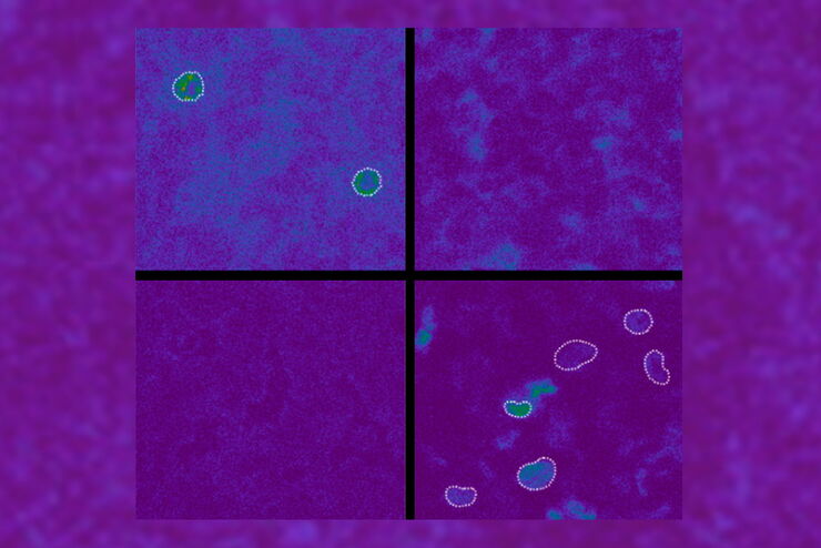
Spectroscopic Evaluation of Red Blood Cells
Hemoglobinopathies are a major healthcare problem. This study presents a possible diagnostic tool for thalassemia which is based on confocal spectroscopy. This approach exploits spectral detection and…

How to Improve Live Cell Imaging with Coral Life
For live-cell CLEM applications, light microscopy imaging is a critical step for identifying the right cell in the right state at the right time. In this article, Leica experts share their insights on…

The Cryo-CLEM Journey
This article describes the Cryo-CLEM technology and the benefits it can provide for scientists. Additionally, some scientific publications are highlighted.
Recent developments in cryo electron…

How to Keep Your Samples Under Physiological Conditions
The Coral Life workflow combines dynamic data with the best possible sample fixation by high pressure freezing. However, good sample preservation won’t help if your cells are stressed by temperature…

Optimizing THUNDER Platform for High-Content Slide Scanning
With rising demand for full-tissue imaging and the need for FL signal quantitation in diverse biological specimens, the limits on HC imaging technology are tested, while user trainability and…

Physiology Image Gallery
Physiology is about the processes and functions within a living organism. Research in physiology focuses on the activities and functions of an organism’s organs, tissues, or cells, including the…

Tissue Image Gallery
Visual analysis of animal and human tissues is critical to understand complex diseases such as cancer or neurodegeneration. From basic immunohistochemistry to intravital imaging, confocal microscopy…
