
Webinar V2 Dark Theme Short
Learn how to use AI to extend live cell imaging in embryo development
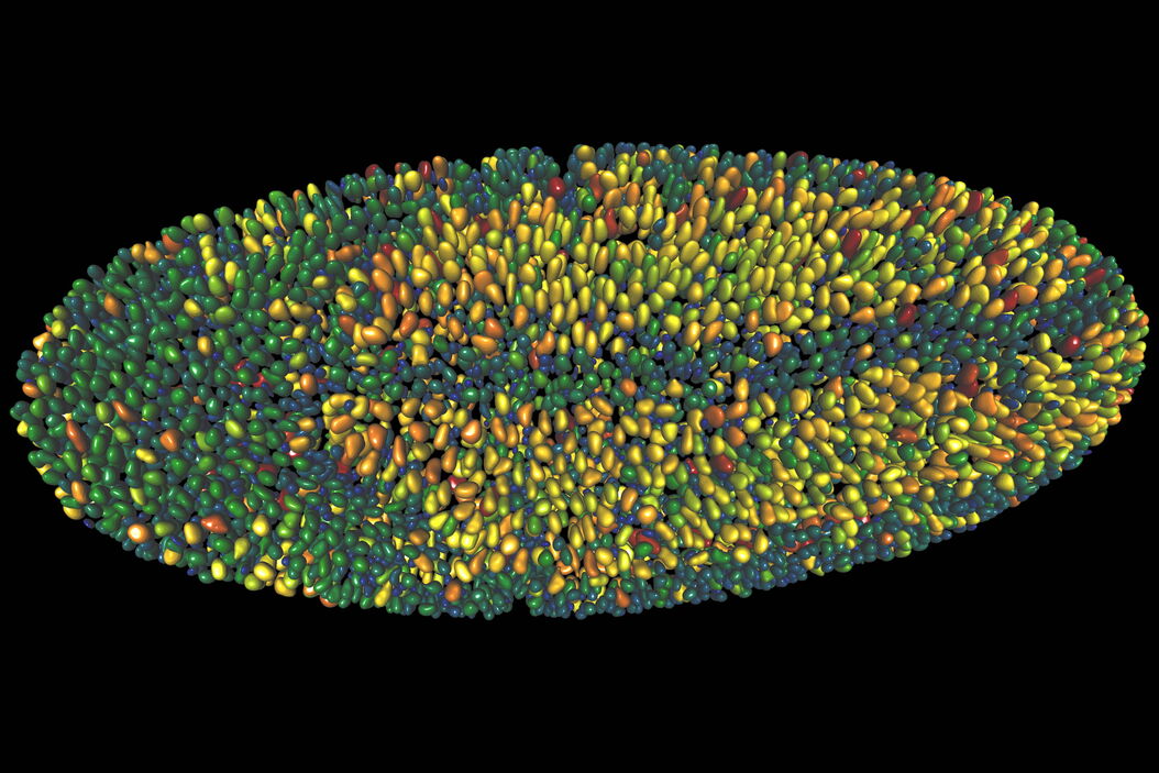
Extended live cell imaging of embryo development requires a delicate balance between light exposure, temporal resolution and spatial resolution to maintain cells’ viability. Compromises between the three factors are needed to achieve the optimal analysis outcome and to unlock greater insights from your imaging data. In this workshop, the Aivia team will demonstrate how AI can help you extend live cell imaging in embryo development.
What will you learn?
Researchers dealing with sample integrity challenges in developmental research can benefit from automated workflows that streamline image analysis.
- Examine critical developmental events in high-definition with minimal sample damage
- Sidestep light exposure and resolution limits to prolong time lapse imaging
- Deploy smart segmentation to easily detect objects
