Aufrechte Lichtmikroskope

Aufrechte Mikroskope für die Life Science-Forschung

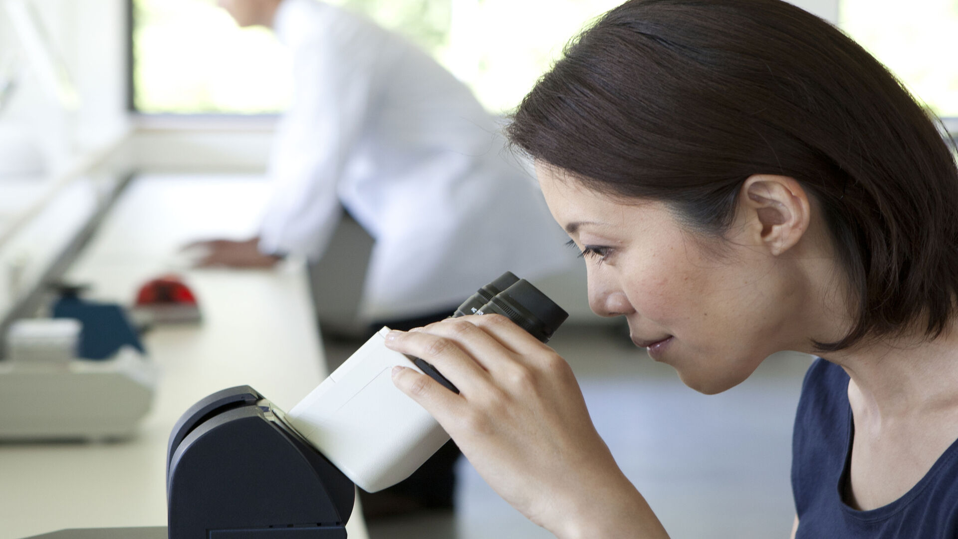
Mit Leica Microsystems erhalten Sie die für Ihre Life Science-Forschung benötigte, hochqualitative Bildgebung und anpassbare aufrechte Mikroskoplösung. Diese leistungsstarken Bildverarbeitungssysteme zeichnen sich durch konstante Farbe, natürliche Lichtverhältnisse, überlegene Optik und konfigurierbare Optionen für kontrastreiche, brillante Bilder für Ihre hochmoderne biologische Forschung aus.

Aufrechte Mikroskope für Industrie und Materialanalyse

Gewinnen Sie entscheidende Erkenntnisse! Mit den aufrechten Mikroskopen von Leica Microsystems für Industrie und Materialanalyse können Sie auch kleinste Details effizient begutachten und prüfen. Damit wir Ihre Anforderungen bestmöglich erfüllen können, bieten wir hervorragende Optik, brillante, kühle LED-Beleuchtung, ergonomisches Zubehör, ausgefeilte Digitalkameras und intuitive Software.

Können wir Ihnen weiterhelfen?

Unsere Spezialisten beantworten gerne Ihre Fragen oder senden Ihnen Informationsmaterial.
Verwandte artikel

Lesen Sie unsere neuesten Artikel

Das Wissensportal von Leica Microsystems bietet Ihnen Wissens- und Lehrmaterial zu den Themen der Mikroskopie. Die Inhalte sind so konzipiert, dass sie Einsteiger, erfahrene Praktiker und Wissenschaftler gleichermaßen bei ihrem alltäglichen Vorgehen und Experimenten unterstützen.

Alle artikel

A fruit fly (Drosophila melanogaster) observed with an Ivesta 3 stereo microscope during fly pushing (sorting of the flies). The scale bar length is 1 mm. Image courtesy of M. Benton, EMBL, Heidelberg, Germany.

A Guide to Using Microscopy for Drosophila (Fruit Fly) Research

The fruit fly, typically Drosophila melanogaster, has been used as a model organism for over a century. One reason is that many disease-related genes are shared between Drosophila and humans. It is…
These images illustrate the need for multiple z-slices to capture all gH2Ax foci in a given cell and get an accurate count.

Development and Derisking of CRISPR Therapies for Rare Diseases

This on-demand presentation by Dr. Fyodor Urnov and Dr. Sadik Kassim, originally delivered at ASGCT 2025, focused on a critical challenge in genetic medicine: how to scale CRISPR therapies from…
Example of calibrating a microscope at a higher magnification value using a stage micrometer.

Microscope Calibration for Measurements: Why and How You Should Do It

Microscope calibration ensures accurate and consistent measurements for inspection, quality control (QC), failure analysis, and research and development (R&D). Calibration steps are described in this…

Integrated Serial Sectioning and Cryo-EM Workflows for 3D Biological Imaging

This on-demand webinar explores how integrated tools can support electron microscopy workflows from sample preparation to image analysis. Experts Andreia Pinto, Adrian Boey, and Hoyin Lai present the…
SEM image of the full Li-NMC electrode sample, showing the two porous layers and the metal film at the center of the structure.

Revealing Sodium Battery Degradation via Cryo-EM and CryoFIB

Explore how cryogenic electron microscopy and focused ion beam techniques uncover the intrinsic structure of sodium battery interfaces. This webinar presents a new degradation model based on separator…
Cell DIVE multiplexed image of FFPE tissue section from human colon adenocarcinoma tissue.

Multiplexed Imaging Reveals Tumor Immune Landscape in Colon Cancer

Cancer immunotherapy benefits few due to resistance and relapse, and combinatorial therapeutic strategies that target multiple steps of the cancer-immunity cycle may improve outcomes. This study shows…
Cell DIVE multiplexed image of FFPE tissue section from human invasive ductal carcinoma (IDC)

AI-Powered Hi-Plex Spatial Analysis Tools for Breast Cancer Research

Breast cancer (BC) is the leading cause of cancer-related deaths in women. Investigating the tumor microenvironment (TME) is crucial to elucidate the mechanisms of tumor progression. Systematic…
Mouse brain slice which was immunostained with GFAP-A647 and imaged using a THUNDER Imager Tissue. Courtesy of H. Xu, University of Pennsylvania, Philadelphia, USA.

Neurowissenschaften

Arbeiten Sie an einem besseren Verständnis neurodegenerativer Erkrankungen oder an einer Untersuchung der Funktionen des Nervensystems? Erfahren Sie, wie Sie mit Bildgebungslösungen von Leica…
Mouse hippocampus brain slice on a grid after HPF using the “Waffle Method”.

The “Waffle Method”: High-Pressure Freeze Complex Samples

This article describes the advantages of a special high pressure freezing method, the so-called “Waffle Method”. Learn how the “Waffle Method” uses EM grids as spacers for high-pressure freezing,…
UC Enuity with Diatome diamond knife.

Mastering Polymer Sectioning with Helmut Gnaegi

When it comes to ultramicrotomy, few names carry the weight of Helmut Gnaegi. As co-founder of Diatome, a global leader in diamond knife technology, Helmut has spent decades refining the art and…

Materialwissenschaft und Inspektion

Optimale Probenvorbereitung und Bildgebungsverfahren sind der Schlüssel für die zuverlässige und genaue Untersuchung von Materialien und die Visualisierung feiner Details. Mit inversen Mikroskopen können Sie dieses Ziel im Vergleich zu aufrechten Mikroskopen auf effizientere Weise erreichen. Sie bieten Ihnen hochwertige Optik und intelligente Automatisierung für optimale Arbeitsabläufe.

Mehr

Biowissenschaftliche Forschung

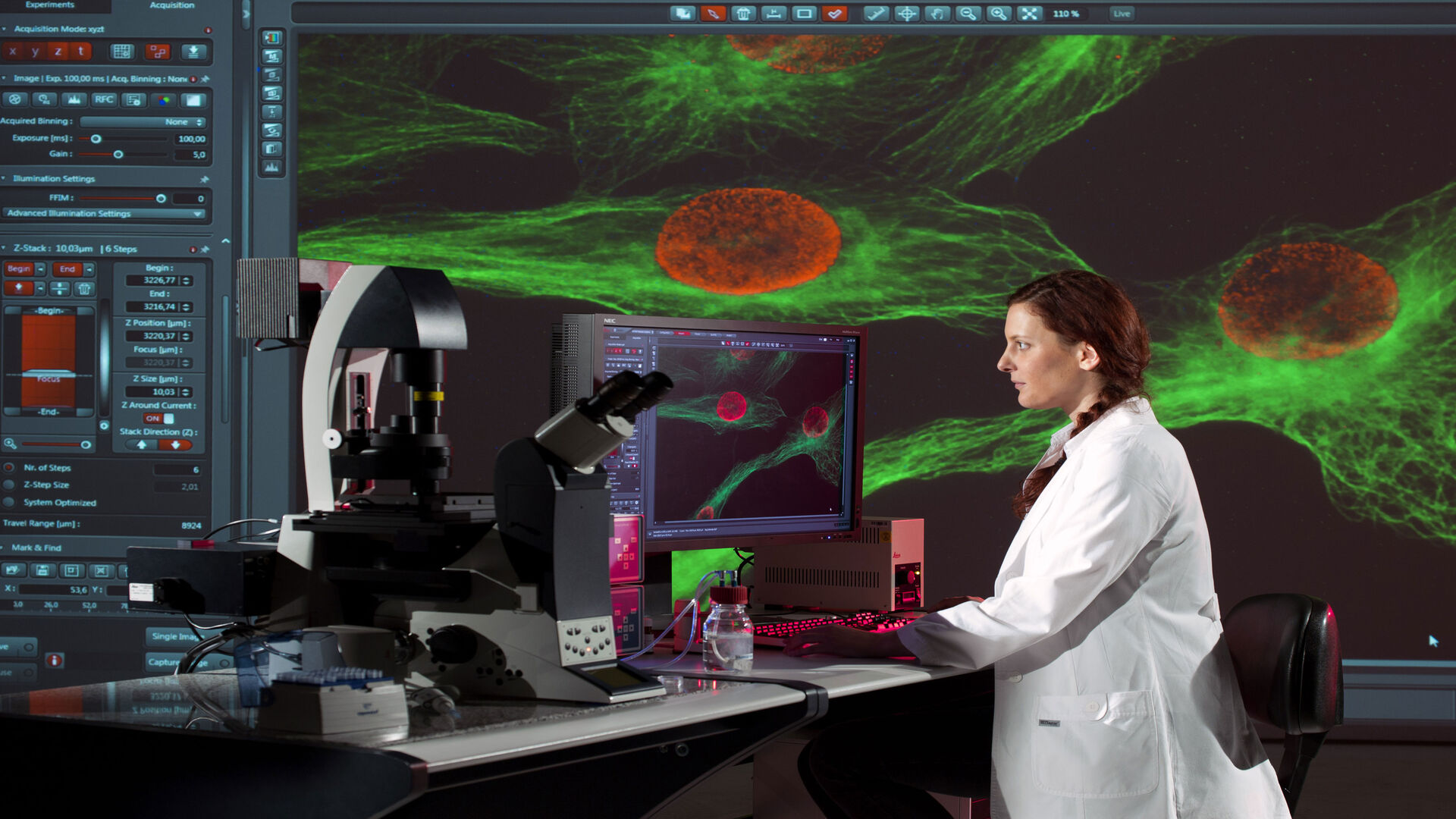
Invertierte Mikroskope werden in der biowissenschaftlichen Forschung häufig für die Visualisierung, Messung, Analyse und Dokumentation von lebenden Zell- und Gewebekulturen eingesetzt. Kontinuierliche innovative technische Fortschritte bei inversen Mikroskopen unterstützen die sich ständig ändernden Bedürfnisse der Biowissenschaftler bei der Bildgebung. 

Mehr

Scroll to top