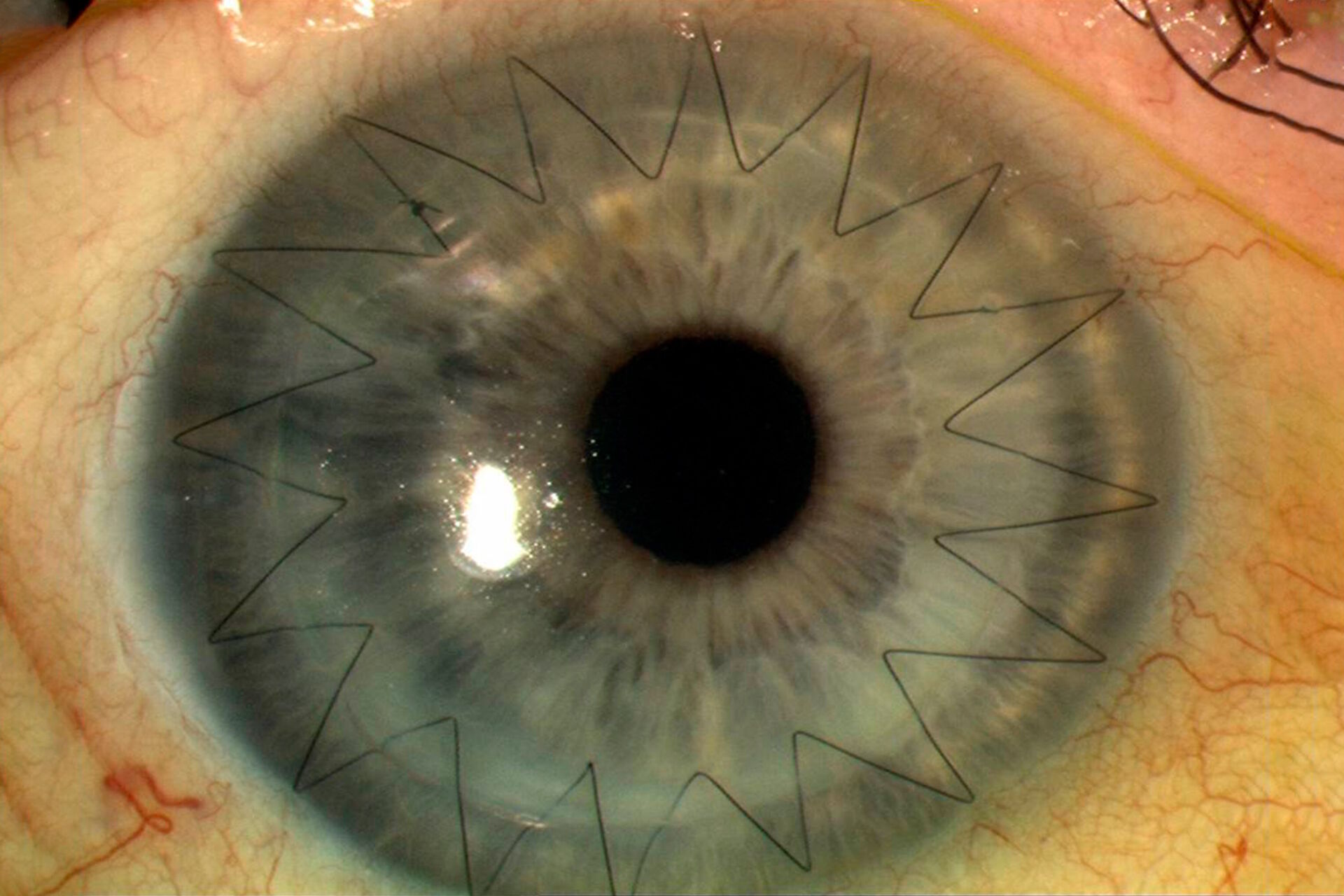
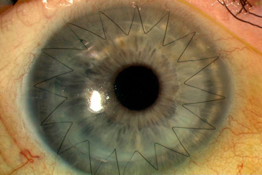
Gain insights from our clinical case study
The use of intraoperative OCT can help overcome the challenges of endothelial keratoplasty through real-time information supporting decision-making. Endothelial keratoplasty is a surgical procedure to replace a damaged endothelium with healthy donor tissue, which includes both DSEK and DMEK techniques.
Key Learnings
- Learn about corneal transplant surgical procedures, including endothelial keratoplasty and partial-thickness corneal transplant
- Discover three clinical cases: Thin Manual Descemet Stripping Endothelial Keratoplasty (TM-DSEK), big-bubble Deep Anterior Lamellar Keratoplasty (DALK) and Descemet Membrane Endothelial Keratoplasty (DMEK)
- Understand the role of intraoperative OCT, in particular for the successful orientation and positioning of the donor tissue in DSEK, DALK and DMEK procedures.
About the author

About Mr. David Anderson
David Anderson is a Consultant Ophthalmic Surgeon and a cornea, cataract and refractive surgery specialist. He has jointly led the corneal fellowship program at Southampton for more than a decade teaching lamellar corneal transplant and anterior segment surgery and has a special interest in refractive surgery including SMILE and topography-guided laser vision correction.
First Clinical Case: Thin Manual Descemet Stripping Endothelial Keratoplasty (TM-DSEK)
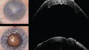
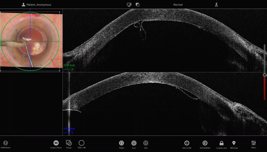
In this case, a Descemet rhexis was created. The clear red reflex on the Proveo 8 with EnFocus intraoperative OCT allowed clear visualization of Descemet’s membrane peeling and endothelium.
When performing DSEK in eyes that have had a penetrating keratoplasty or other pathologies such as posterior membrane dystrophies, it is valuable to see DM as opposed to stromal tears or folds.
In TM-DSEK, it is possible to leave some of the DM present whereas in DMEK, DM is stripped beyond the border of the graft.
When performing the strip, the intraoperative OCT provided live confirmation of where DM was present. This is particularly helpful when there is a poor view e.g. in this case of ocular trauma. The OCT showed the separation of Descemet’s membrane very clearly.
Continue Reading
Learn more about this case and discover two other corneal transplantation cases: Big-bubble Deep Anterior Lamellar Keratoplasty (DALK) and Descemet Membrane Endothelial Keratoplasty (DMEK).
Please note that off-label uses of products may be discussed. Please check with regulatory affairs for cleared indications for use in your region. The statements of the healthcare professionals included in this clinical case reflect only their opinion and personal experience and not those of Leica Microsystems. They also do not necessarily reflect the opinion of any institution with whom they are affiliated.
