Contact Us

Intraoperative OCT-Assisted Corneal Transplant Procedures

Discover how intraoperative OCT supports cornea surgeons with enhanced visualization during corneal transplantation and can facilitate the positioning and adaptation of the donor cornea

Keratoplasty of pathologic cornea Pathologic_cornea.jpg

Penetrating keratoplasty, also known as corneal transplant, is the surgical removal of a damaged or diseased portion of the cornea, followed by the implantation of a healthy donor cornea.

Several intraoperative complications may occur during corneal transplant surgery including poor graft centration, irregular trephination, damage to the lens, damage to the donor tissue, choroidal hemorrhage and effusion, incarceration of iris tissue in the wound and vitreous in the anterior chamber [1]​​​​​​​.

About the Clinical Case Study

Key Learnings

  •  Learn about corneal transplant surgery and its challenges. 
  • Discover three corneal transplant surgery clinical cases and how to overcome challenges. 
  • Understand the role of intraoperative OCT, in particular for the successful positioning and adaptation of the donor cornea.

The use of intraoperative optical coherence tomography (OCT) provides anterior segment surgeons with enhanced visualization during corneal transplantation contributing to better results and increased safety. Prof. Nikolaos Bechrakis demonstrates its value through three clinical cases. 

First Clinical Case: Bullous Keratopathy

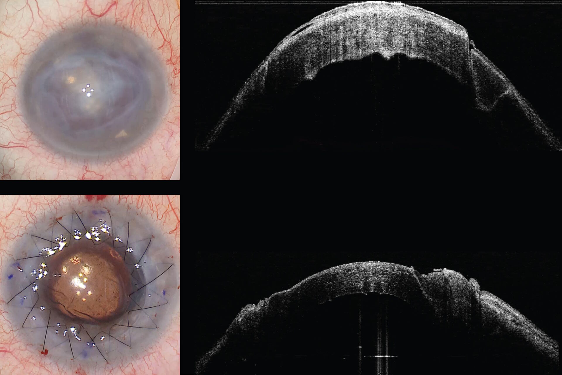
In this clinical case, the patient had an extremely pathologic cornea, with the epithelium showing bullous keratopathy and a very irregular posterior surface of the cornea. The image from the EnFocus intraoperative OCT indicated the area where previous surgery had been performed on the cornea and where the cornea was trephined for the new keratoplasty. 

The intraoperative OCT showed how the viscoelastic was filling the anterior chamber. It allowed Prof. Bechrakis to see exactly where the previous scars were to avoid the area where the previous trephination had occurred.

Continue Reading

Learn more about this case and discover two more corneal transplantation cases.

Enter your information to download the full case study.  
 

READ THE CASE STUDY

By clicking on the SUBMIT button, I confirm that I have reviewed and agree with Leica Microsystems GmbH Terms of Use and their Privacy Policy. I also understand my privacy choices as they pertain to my personal data, as detailed in the aforementioned Privacy Policy under ‘’Your Privacy Choices’’.

Please note that off-label uses of products may be discussed. Please check with regulatory affairs for cleared indications for use in your region. The statements of the healthcare professionals included in this clinical case reflect only their opinion and personal experience and not those of Leica Microsystems. They also do not necessarily reflect the opinion of any institution with whom they are affiliated.

Related Articles

Background image
Scroll to top