Leica M530 OHX
Microscopi operatori
Prodotti
Home
Leica Microsystems
Leica M530 OHX Microscopio per microchirurgia
Per rimanere focalizzati
Leggi gli articoli più recenti
Launching a Neurosurgical Department with Limited Resources
Learn about Dr. Claire Karekezi’s journey and experience launching a neurosurgical department within the Rwanda Military Hospital with limited resources.
Use of AR Fluorescence in Neurovascular Surgery
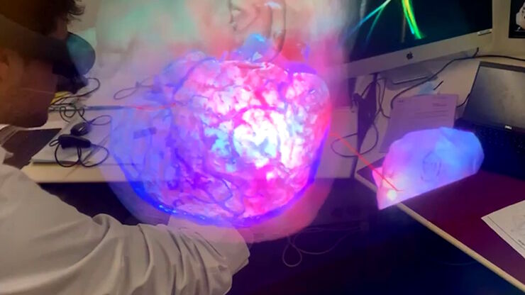
Learn about the use of GLOW800 Augmented Reality in neurovascular surgery through clinical cases and videos, including aneurysm and tumor resection cases.
3D, AR & VR for Teaching in Neurosurgery
Discover the evolution of neurosurgical teaching and how 3D, Augmented Reality and Virtual Reality can help better learn anatomy and acquire surgical skills.
Surgical Management of High-Grade Gliomas
Learn about the surgical management of high-grade gliomas and how to expand the extent of resection intra-operatively using tools such as 5-ALA fluorescence.
Neurosurgical Treatment of Spinal Arterio-Venous Fistulas
Learn about the neurosurgical treatment of spinal arterio-venous fistulas, including classification, epidemiology and surgical approaches.
Skull Base Neurosurgery: Epidural Lateral Approaches
Surgery of skull base tumors and diseases, such as cavernomas, epidermoid cysts, meningiomas and schwannomas, can be quite complex. During the Leica 2021 Neurovisualization Summit, a unique event…
Using GLOW800 AR in Radial Forearm Flap Free Phalloplasty
In this video, Chief Microsurgeon Professor Küntscher and his team perform a radial forearm free flap phalloplasty and use ICG fluorescence imaging to show the blood flow in the whole flap from the…
How AR Surgery Benefits Radial Forearm Free Flap Phalloplasty
The goal of penile reconstruction is to provide an aesthetic penoid with tactile and erogenous sensation, so the patient can have sexual intercourse and void standing.1 Currently, the radial forearm…
Free Flap Procedures in Oncological Reconstructive Surgery
Free flap surgery is considered the gold standard for breast, head and neck reconstructions for cancer patients. These procedures, which enable functional and aesthetic rehabilitation, can be quite…
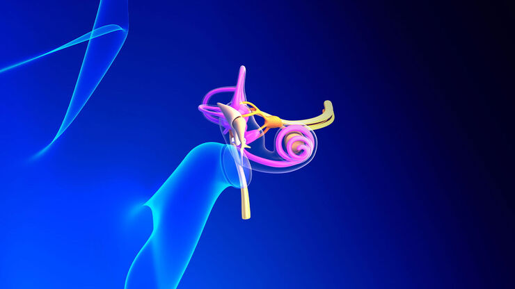
Minor’s Syndrome Surgical Intervention by Prof. Vincent Darrouzet
Minor’s disease, also called Superior Semicircular Canal Dehiscence (SSCD) or Minor’s syndrome, is a rare disorder of the inner ear that affects hearing and balance. The disease is characterized by…
How to Choose a Microscope for Reconstructive Surgery
Plastic and reconstructive surgery requires excellent visualization to repair intricate and fine structures. Oncological reconstructive surgery procedures are among the most delicate, including breast…
Advances in Oncological Reconstructive Surgery
Decision making and patient care in oncological reconstructive surgery have considerably evolved in recent years. New surgical assistance technologies are helping surgeons push the boundaries of what…
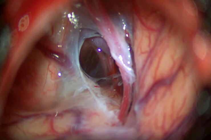
Optimal Visualization in Brain Surgery
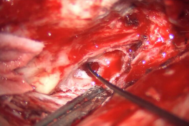
This case study “Treatment of the Galassi type III arachnoid cyst with the M530 OHX surgical microscope from Leica Microsystems” documents the procedure step by step and shows the visualization…
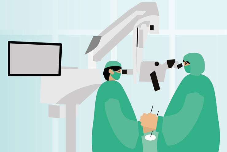
How to use a Surgical Microscope as an Operating Room Nurse
Surgical microscopes play an essential role in the modern microsurgery procedures. It provides the surgeon, assistant and operating room staff with a magnified and illuminated high-quality image of…
How Augmented Reality is Transforming Vascular Neurosurgery
Augmented Reality is changing surgery, with new information helping to improve the precision and safety of procedures. This is especially true in vascular neurosurgery where Augmented Reality is…
Neurovascular Surgery & Augmented Reality Fluorescence
Vascular neurosurgery is highly complex. Surgeons need to be able to rely on robust anatomical information. As such, visualization technologies play an essential role.
Prof. Nils Ole Schmidt is a…
Oncological Reconstructive Surgery with the Leica M530 OHX Microscope
Precision is essential in oncological reconstructive surgery, in particular when it relies on free flap techniques. Microsurgical microscopes provide optimal visualization and help streamline the…
Oncological Reconstructive Surgery: Why Use a Microscope
Recent advances in microsurgery are enhancing breast reconstruction for oncology patients, allowing both functional and aesthetic rehabilitation. More and more surgeons are adopting surgical…
Plastic & Reconstructive Surgery: Why Use a Microscope
Plastic and Reconstructive Surgery procedures can be delicate. Visualization solutions play an essential role, allowing to perform the surgery in the best conditions. And more and more plastic…
Minimally Invasive Spine Surgery: Improving Precision and Accuracy with Microscopes
Spine surgery is extremely delicate and requires extensive training and experience. Innovative visualization technologies can also help achieve better outcomes allowing to see more and have a clearer…
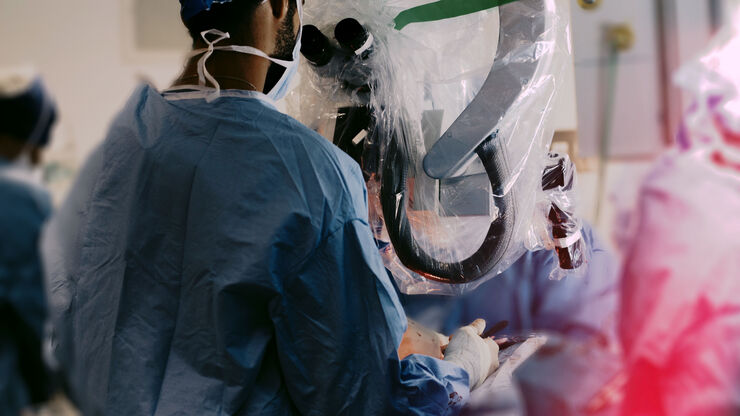
Surgical Microscopes: Key Factors for OR Nurses
Operating room (OR) nurses are vital to the surgery process. An OR Nurse Manager explains the key surgical microscope features to facilitate their work.
How to Drape an Overhead Surgical Microscope
The tutorial features the Leica ARveo digital Augmented Reality microscope for complex neurosurgery. The procedure also applies to the Leica M530 OHX, OH6, OH5 and OH4.
FusionOptics in Neurosurgery and Ophthalmology – for a Larger 3D Area in Focus
Neurosurgeons and ophthalmologists deal with delicate structures, deep or narow cavities and tiny structures with vitally important functions. A clear, three-dimensional view on the surgical field is…
Fields of Application
Neurochirurgia
I microscopi per neurochirurgia di Leica Microsystems offrono ottiche e ingegneria di alta qualità per chirurgia cranica, spinale, multidisciplinare e per altre discipline di microchirurgia.
Chirurgia neurovascolare
Esplorate i microscopi chirurgici di Leica Microsystems per la chirurgia neurovascolare, potenziati con applicazioni di imaging a fluorescenza e fluorescenza AR. Ottenete informazioni critiche…


