
The benefits of 3D for neurosurgical teaching
A study conducted with medical students1 has shown that they see many benefits to the use of 3D for anatomical education. 88% believe it allows them to better learn and understand anatomy and 85.7% think the use of 3D tutorial videos should be integrated into routine teaching. However, the majority of students prefer cadaver dissections to virtual visualizations. As such, 3D is a complementary tool. The same study has demonstrated that learning is better assimilated at 1 month with 3D vs 2D, in particular for spatial relationship understanding and clinical inference.
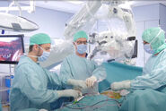
The use of 3D is also valuable from a teacher perspective, Dr. Bernard says, helping to share experience and knowledge with a large number of students. GLOW 3D from Leica Microsystems allows to create and visualize different kinds of 3D content including Augmented Reality, Virtual Reality and 3D videos. Neurosurgeons can easily record 3D content during surgical procedures and build video tutorials to be watched on headsets or screens. This allows students to see what the neurosurgeon sees in the microscope eyepieces during surgery.
How to Develop an Educational Surgery Video
According to Dr. Bernard, to develop an educational surgery video, the first step is to record the surgery, which can be done with the Leica M530 OHX surgical microscope. Then, the creation of a script enables to build the story flow and integrate different perspectives. For example, a video developed as part of a French Neurosurgery Society workshop for residents on combined petrosectomy included advice from both the ENT surgeon and neurosurgeons. It is advisable to have a professional audio recording. Text can also be included in the video to provide additional background.
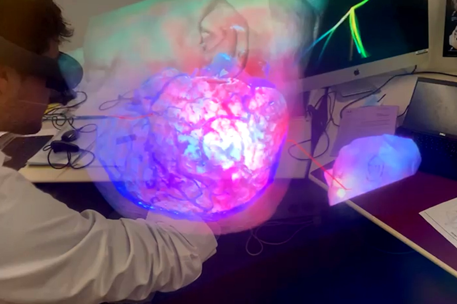
Educational surgery videos can be shared through Virtual Reality headsets and 3D screens. They can also be published in scientific journals. Operative Neurosurgery published the combined petrosectomy stereoscopic surgical video produced by Dr. Bernard and colleagues.

This video was used in a French Neurosurgery Society workshop and received very positive student feedback. Students thought there were many valuable aspects including the ease of use, immersiveness, the ability to go back and repeat as well the ability to get tips and tricks. This type of tool can support a better mental representation, leading to better technical skills.
To create and promote 3D content, Dr. Bernard has participated in the creation of a 3D Lab within the University of Angers. He has also launched a YouTube channel on Anatomy with 3D videos and participated in the creation of a Virtual Reality application for medical students called Akivi (Anatomical Knowledge in Virtual Immersion).

New technologies such as Augmented Reality and Virtual Reality are not only valuable for medical students. They can also help residents and young neurosurgeons improve their practice, for example by developing their surgical planning skills. In the future, these technologies could also enhance surgical research.
Want to learn more? Watch the full webinar presented by Dr. Florian Bernard.
Watch the video on YouTube
Disclaimer: “Please note that off-label uses of products may be discussed. Please check with regulatory affairs for cleared indications for use in your region. The statements of the healthcare professionals included in this symposium reflect only their opinion and personal experience and not those of Leica Microsystems. They also do not necessarily reflect the opinion of any institution with whom they are affiliated”
 case.")