Aufrechte Lichtmikroskope

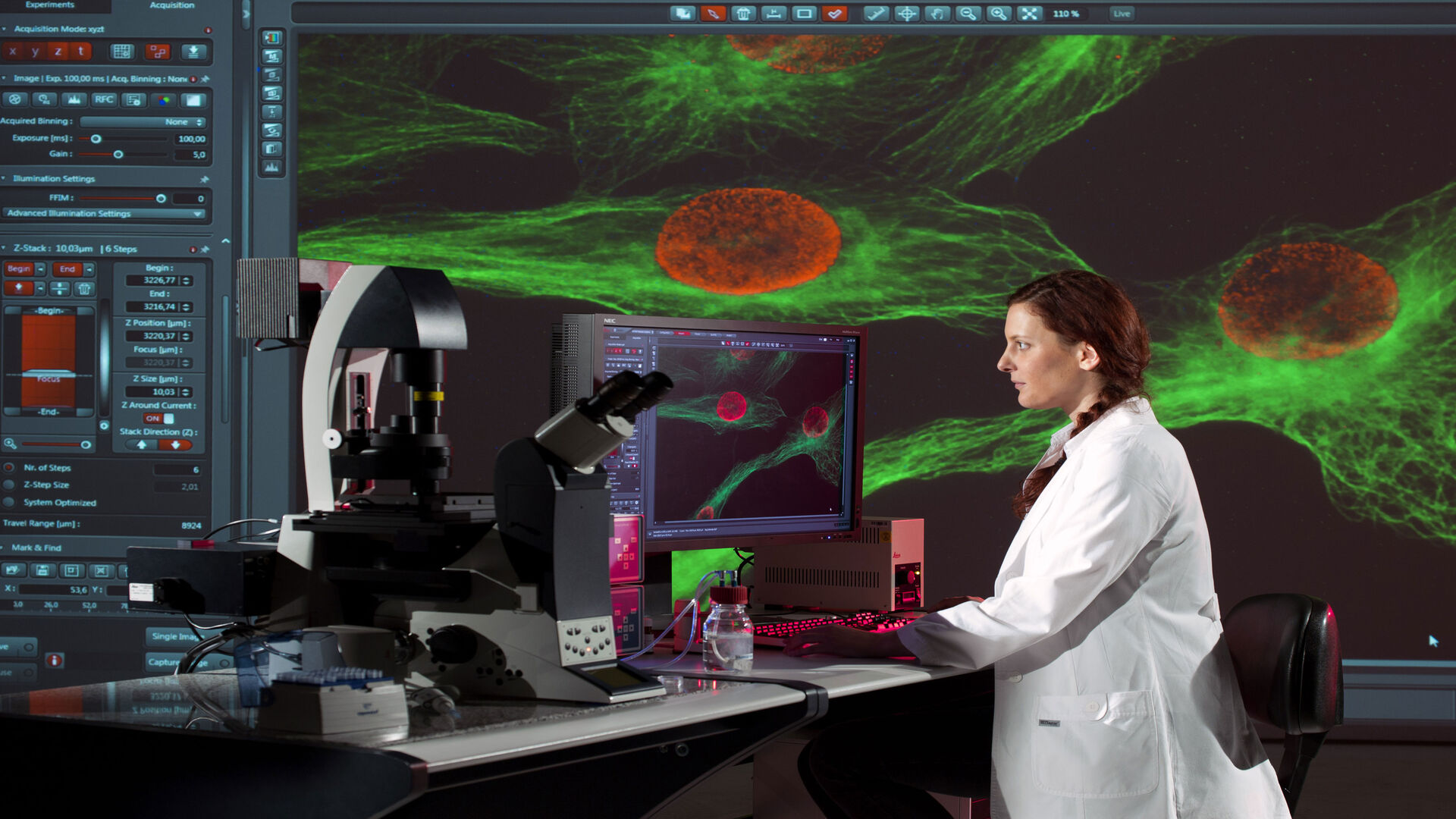
Aufrechte Mikroskope für die Life Science-Forschung

Mit Leica Microsystems erhalten Sie die für Ihre Life Science-Forschung benötigte, hochqualitative Bildgebung und anpassbare aufrechte Mikroskoplösung. Diese leistungsstarken Bildverarbeitungssysteme zeichnen sich durch konstante Farbe, natürliche Lichtverhältnisse, überlegene Optik und konfigurierbare Optionen für kontrastreiche, brillante Bilder für Ihre hochmoderne biologische Forschung aus.

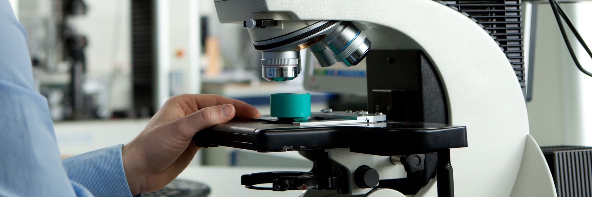
Aufrechte Mikroskope für Industrie und Materialanalyse

Gewinnen Sie entscheidende Erkenntnisse! Mit den aufrechten Mikroskopen von Leica Microsystems für Industrie und Materialanalyse können Sie auch kleinste Details effizient begutachten und prüfen. Damit wir Ihre Anforderungen bestmöglich erfüllen können, bieten wir hervorragende Optik, brillante, kühle LED-Beleuchtung, ergonomisches Zubehör, ausgefeilte Digitalkameras und intuitive Software.

Können wir Ihnen weiterhelfen?

Unsere Spezialisten beantworten gerne Ihre Fragen oder senden Ihnen Informationsmaterial.

Verwandte artikel

Lesen Sie unsere neuesten Artikel

Das Wissensportal von Leica Microsystems bietet Ihnen Wissens- und Lehrmaterial zu den Themen der Mikroskopie. Die Inhalte sind so konzipiert, dass sie Einsteiger, erfahrene Praktiker und Wissenschaftler gleichermaßen bei ihrem alltäglichen Vorgehen und Experimenten unterstützen.

Alle artikel

Final Segmentation of organelles in Trichomonas species. Magenta – costa, light blue – hydrogenosomes, turquoise – ER, red – vacuoles, yellow – axostyle, green – Golgi apparatus.  Sample courtesy of Isabelle Guerin-Bonne, Low Kay En, Electron Microscopy Unit, Yong Loo Lin School of Medicine, National University of Singapore. Scale bar: 1 µm.

Volume EM and AI Image Analysis

The article outlines a detailed workflow for studying biological tissues in three dimensions using volume-scanning electron microscopy (volume-SEM) combined with AI-assisted image analysis. The focus…
Image of roundworm C. elegans acquired with a M205 FA fluorescence automated stereo microscope in combination with Rottermann contrast. Areas labelled with mCherry are seen as reddish purple.

A Guide to C. elegans Research – Working with Nematodes

Efficient microscopy techniques for C. elegans research are outlined in this guide. As a widely used model organism with about 70% gene homology to humans, the nematode Caenorhabditis elegans (also…
Dr. Nordmann in conversation with Dr. Falk Schlaudraff, Manager Product Management Uprights (widefield/compound) at Leica Microsystems

How a Breakthrough in Spatial Proteomics Saved Lives

Toxic epidermal necrolysis (TEN) is a rare but devastating reaction to common medications like antibiotics or gout treatments. It begins innocuously, often as a rash, but can escalate rapidly into…
Dr. Andrew Huang, Baylor College of Medicine, in the operating room (OR) performing ear, nose and throat (ENT) surgery using the MyVeo surgical visualization headset.

A Microvascular Surgeon’s View: How MyVeo Transforms Visualization

In this article, Dr. Andrew T. Huang, MD, FACS, otolaryngologist and a head and neck reconstructive surgeon, shares how digital 3D surgical visualization with the MyVeo headset from Leica Microsystems…

A Novel Laser-Based Method for Studying Optic Nerve Regeneration

Optic nerve regeneration is a major challenge in neurobiology due to the limited self-repair capacity of the mammalian central nervous system (CNS) and the inconsistency of traditional injury models.…
Fluorescence microscopy of sectioned tissue, showing the interface between the extensor digitorum longus muscle and the common peroneal nerve in the adult rat. Regenerative peripheral nerve interface (RPNI) at 2 weeks. Image acquired using Mica. Stained for nuclei (blue), neurofilaments (green) and S100B (red). Image courtesy of Dr. Aaron Lee, Department of Bioengineering (Lab of Dr. Rylie Green), Imperial College London.

How to Image Axon Regeneration in Deep Muscle Tissue

This study highlights Dr. Aaron Lee’s research on mapping nerve regeneration in muscle grafts post-amputation. Limb loss often leads to reduced quality of life, not only from tissue loss but also due…
5 hour time-lapse maximum intensity projection of a zebrafish embryo along the z-axis at 3 days post fertilization. Left: microglia cells. Right: bright field channel. Courtesy of Prof. Francesca Peri, University of Zurich, Switzerland.

Capturing Developmental Dynamics in 3D

This application note showcases how the Viventis Deep dual-view light sheet microscope was successfully used by researchers for exploring high-resolution, long-term imaging of 3D multicellular models…
Some 2D measurements, e.g., lengths and areas, made on a PCB sample with a Leica measurement microscope using the Enersight software.

How to Select the Right Measurement Microscope

With a measurement microscope, users can measure the size and dimensions of sample features in both 2D and 3D, something crucial for inspection, QC, failure analysis, and R&D. However, choosing the…
A fruit fly (Drosophila melanogaster) observed with an Ivesta 3 stereo microscope during fly pushing (sorting of the flies). The scale bar length is 1 mm. Image courtesy of M. Benton, EMBL, Heidelberg, Germany.

A Guide to Using Microscopy for Drosophila (Fruit Fly) Research

The fruit fly, typically Drosophila melanogaster, has been used as a model organism for over a century. One reason is that many disease-related genes are shared between Drosophila and humans. It is…
These images illustrate the need for multiple z-slices to capture all gH2Ax foci in a given cell and get an accurate count.

Development and Derisking of CRISPR Therapies for Rare Diseases

This on-demand presentation by Dr. Fyodor Urnov and Dr. Sadik Kassim, originally delivered at ASGCT 2025, focused on a critical challenge in genetic medicine: how to scale CRISPR therapies from…

Materialwissenschaft und Inspektion

Optimale Probenvorbereitung und Bildgebungsverfahren sind der Schlüssel für die zuverlässige und genaue Untersuchung von Materialien und die Visualisierung feiner Details. Mit inversen Mikroskopen können Sie dieses Ziel im Vergleich zu aufrechten Mikroskopen auf effizientere Weise erreichen. Sie bieten Ihnen hochwertige Optik und intelligente Automatisierung für optimale Arbeitsabläufe.

Erfahren Sie mehr

Biowissenschaftliche Forschung

Invertierte Mikroskope werden in der biowissenschaftlichen Forschung häufig für die Visualisierung, Messung, Analyse und Dokumentation von lebenden Zell- und Gewebekulturen eingesetzt. Kontinuierliche innovative technische Fortschritte bei inversen Mikroskopen unterstützen die sich ständig ändernden Bedürfnisse der Biowissenschaftler bei der Bildgebung. 

Erfahren Sie mehr

Scroll to top