EM ICE
Elektronenmikroskopie Probenvorbereitung
Produkte
Startseite
Leica Microsystems
EM ICE Hochdruckgefriersystem
Halten Sie den Augenblick Ihrer Entdeckung fest
Lesen Sie unsere neuesten Artikel
, insulin SGs (orange), microtubules (red), nucleus (yellow), and plasma membrane (transparent).")
High-Pressure Freezing Protocols for Ultrastructural 3D EM
High pressure freezing (HPF) can help preserve hydrated cells and tissues close to their biological state at the moment of immobilization, supporting more reliable ultrastructural interpretation than…
 labeled with membrane-permeable calcein, high-pressure frozen in salt water using EM ICE.")
High-Pressure Freezing for Organoids: Cryo CLEM & FIB Lift Out
Master cryo EM workflow steps for challenging 3D samples: when to choose HPF vs. plunge freezing, reproducible blotting/ice control, contamination aware transfers, Cryo CLEM 3D targeting in organoids,…
 image of a cross section of C. elegans (roundworm). Courtesy of T. Müller-Reichert, MPI-CBG, Dresden, Germany and K. McDonald, University of California, Berkeley, USA.")
Brief Introduction to High-Pressure Freezing for Cryo-Fixation
Preparation of biological specimens for electron microscopy (EM) often requires cryo-fixation which does not introduce significant structural alterations of cellular constituents. A common method used…

Neurowissenschaften
Arbeiten Sie an einem besseren Verständnis neurodegenerativer Erkrankungen oder an einer Untersuchung der Funktionen des Nervensystems? Erfahren Sie, wie Sie mit Bildgebungslösungen von Leica…

The “Waffle Method”: High-Pressure Freeze Complex Samples
This article describes the advantages of a special high pressure freezing method, the so-called “Waffle Method”. Learn how the “Waffle Method” uses EM grids as spacers for high-pressure freezing,…

Wie die Analyse von Meeresmikroorganismen durch Hochdruckgefrieren verbessert werden kann
Die ultrastrukturelle Analyse von Umweltproben, hier Dinoflagellaten, bleibt heutzutage eine Herausforderung. Hier zeigen wir, dass die Durchführung von Hochdruckgefrieren (HPF) vor Ort die…

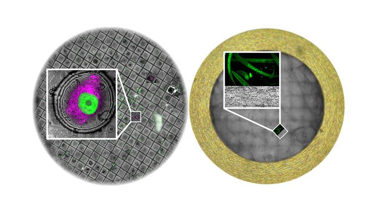
How to Successfully Perform Live-Cell CLEM
The Leica Nano workflow provides a streamlined live-cell CLEM solution for getting insight bout structural changes of cellular components over time. Besides the technical handling described in the…

How to Improve Live Cell Imaging with Coral Life
For live-cell CLEM applications, light microscopy imaging is a critical step for identifying the right cell in the right state at the right time. In this article, Leica experts share their insights on…

How to Keep Your Samples Under Physiological Conditions
The Coral Life workflow combines dynamic data with the best possible sample fixation by high pressure freezing. However, good sample preservation won’t help if your cells are stressed by temperature…

Putting Dynamic Live Cell Data into the Ultrastructural Context
With workflow Coral Life, searching for a needle in the haystack is a thing of the past. Take advantage of correlative light and electron microscopy to identify directly the right cell at the right…

Fast, High-quality Vitrification with the EM ICE High Pressure Freezer
The EM ICE High Pressure Freezer was developed with a unique freezing principle and uses only a single pressurization and cooling liquid: liquified nitrogen (LN2). This design enables three major…

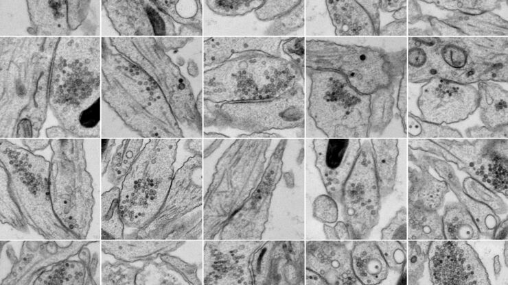
Investigating Synapses in Brain Slices with Enhanced Functional Electron Microscopy
A fundamental question of neuroscience is: what is the relationship between structural and functional properties of synapses? Over the last few decades, electrophysiology has shed light on synaptic…

High-pressure freezing: Revealing functional mechanisms of synaptic transmission
Learn more about applying optogenetic stimulation in the EM ICE and how this technology has the potential to reveal structural and functional mechanisms of synaptic transmission. Get a detailed…
Workflows and Instrumentation for Cryo-electron Microscopy
Cryo-electron microscopy is an increasingly popular modality to study the structures of macromolecular complexes and has enabled numerous new insights in cell biology. In recent years, cryo-electron…

Modellorganismen in der Forschung
Modellorganismen sind Spezies, mit denen Forscher bestimmte biologische Vorgänge untersuchen. Sie haben genetische Ähnlichkeiten mit Menschen und werden häufig in Forschungsbereichen wie Genetik,…

Virologie
Liegt Ihr Forschungsschwerpunkt auf Virusinfektionen und -krankheiten? Erfahren Sie, wie Sie mit Lösungen für Bildgebung und Probenvorbereitung von Leica Microsystems mehr Erkenntnisse in der…

Expert Knowledge on High Pressure Freezing and Freeze Fracturing in the Cryo SEM Workflow
Get an insight in the working methods of the laboratory and learn about the advantages of Cryo SEM investigation in EM Sample Preparation. Find out how high pressure freezing, freeze fracturing and…
Bridging Structure and Dynamics at the Nanoscale through Optogenetics and Electrical Stimulation
Nanoscale ultrastructural information is typically obtained by means of static imaging of a fixed and processed specimen. However, this is only a snapshot of one moment within a dynamic system in…
EM‑Probenvorbereitung: Typische Workflows und Anwendungen
Mit den Probenvorbereitungslösungen von Leica Microsystems können Forscher bei der Abbildung von Proben mit der Elektronenmikroskopie durchgängig hochwertige, präzise und reproduzierbare Ergebnisse…
Anwendungsbereiche
Korrelative Licht- und Elektronenmikroskopie (CLEM)
Coral Workflows von Leica Microsystems unterstützt Nutzer dabei, Daten aus der Fluoreszenz- und Elektronenmikroskopie zu korrelieren (CLEM).
EM‑Probenvorbereitung: Typische Workflows und Anwendungen
Mit den Probenvorbereitungslösungen von Leica Microsystems können Forscher bei der Abbildung von Proben mit der Elektronenmikroskopie durchgängig hochwertige, präzise und reproduzierbare Ergebnisse…
