
Spécialités médicales
Spécialités médicales
Explorez une collection complète de ressources scientifiques et cliniques conçues pour les professionnels de la santé, notamment des points de vue de pairs, des études de cas cliniques et des symposiums. Conçue pour les neurochirurgiens, les ophtalmologues et les spécialistes en chirurgie plastique et reconstructive, en ORL et en dentisterie. Cette collection met en lumière les dernières avancées en matière de microscopie chirurgicale. Découvrez comment les technologies chirurgicales de pointe, telles que la fluorescence AR, la visualisation 3D et l'imagerie OCT peropératoire, permettent de prendre des décisions en toute confiance et d'être précis dans les chirurgies complexes.
Filter articles
Tags
Type de publication
Loading...
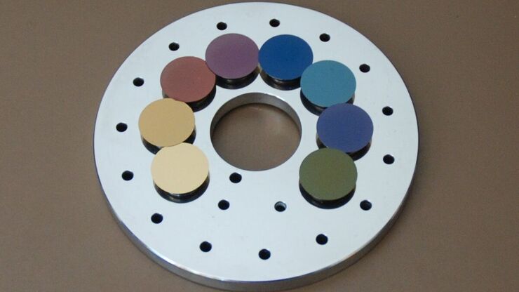
Carbon Thickness Evaluation in Electron Microscopy
The coating layers applied and used for electron microscopy imaging are commonly controlled and measured by quartz crystals. These crystals oscillate with a certain frequency (around 6 megahertz when…
Loading...
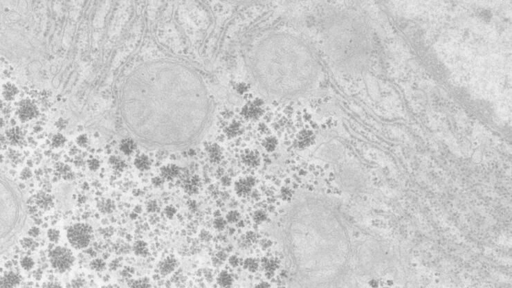
Perusing Alternatives for Automated Staining of TEM Thin Sections
Contrast in transmission electron microscopy (TEM) is mainly produced by electron scattering at the specimen: Structures that strongly scatter electrons are referred to as electron dense and appear as…
Loading...

Brief Introduction to Critical Point Drying
One of the uses of the Scanning Electron Microscope (SEM) is in the study of surface morphology in biological applications which requires the preservation of the surface details of a specimen. Samples…
