Kontaktieren Sie uns

James DeRose , Ph.D.

James DeRose

James DeRose ist Global Marketing Communication Manager bei Leica Microsystems. Sein Schwerpunkt liegt auf der Erstellung optimierter Inhalte für Anwendungen in den Bereichen Life Science, Materialwissenschaften sowie Industrie und Fertigung. Er erstellt Applikationsberichte, Fallstudien, technische Berichte, White Papers, Anwendungsseiten, Produktseiten, Social Media Posts, E-Mails, Testimonials und andere Materialien. Er arbeitet seit 2013 bei Leica Microsystems. In der Vergangenheit arbeitete er an Anwendungsentwicklungsprojekten in den Bereichen Grenzflächenchemie und -physik, Wärme- und Verfahrenstechnik, Korrosion und Metallographie, Oberflächenbeschichtungen, Materialwissenschaften, Biotechnologie und Zellbiologie. Er hat Erfahrung mit verschiedenen Arten von Mikroskopie- und Analysemethoden.

See the Structure with Microscopy - Know the Composition with Laser Spectroscopy

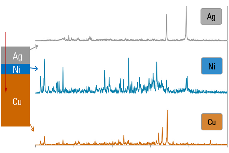
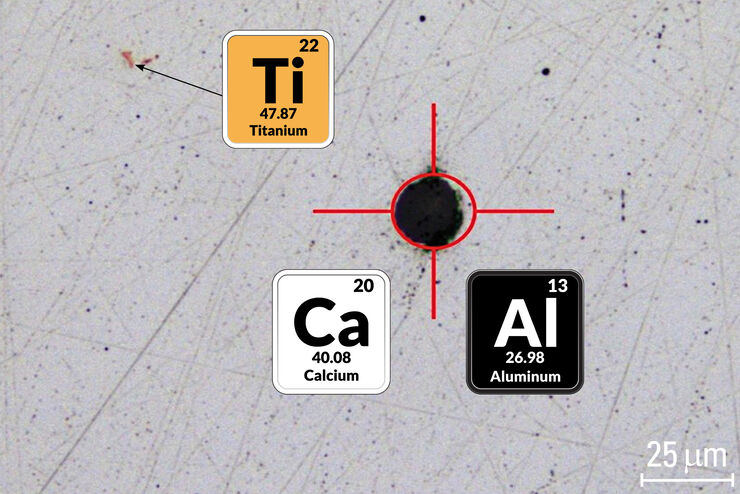
The advantages of a 2-in-1 materials analysis solution combining optical microscopy and laser induced breakdown spectroscopy (LIBS) for simultaneous visual and chemical inspection are described in…
Convalaria

Introduction to Widefield Microscopy

This article gives an introduction to widefield microscopy, one of the most basic and commonly used microscopy techniques. It also shows the basic differences between widefield and confocal…
Comparison when observing a rodent model organism with a Greenough versus CMO (common main objective) stereo microscope for a task like surgery.

Rodent and Small-Animal Surgery

Learn how you can perform rodent (mouse, rat, hamster) and small-animal surgery efficiently with a microscope for developmental biology and medical research applications by reading this article.
Coaxial light with only polarizer open.

Digital Microscopy with Versatile Illumination and Various Contrast Methods for More Efficient Inspection and Quality Control

State-of-the-art digital microscopes utilizing a versatile illumination system capable of achieving multiple contrast methods, such as the Leica DVM6, are very useful for inspection, quality control,…
Transgenic zebrafish larva where fluorescent proteins label the heart muscle blue, blood and blood vessels red, and all circulatory system cells green. Image recorded with a M205 FA microscope.

Imaging and Analyzing Zebrafish, Medaka, and Xenopus

Discover how to image and analyze zebrafish, medaka, and Xenopus frog model organisms efficiently with a microscope for developmental biology applications from this article.
Fluorescence stereo microscope image of anesthetized Mediterranean fruit flies recorded with a M205 stereo microscope.

Investigating Fruit Flies (Drosophila melanogaster)

Learn how to image and investigate Drosophila fruit fly model organisms efficiently with a microscope for developmental biology applications from this article.
C. elegans

Studying Caenorhabditis elegans (C. elegans)

Find out how you can image and study C. elegans roundworm model organisms efficiently with a microscope for developmental biology applications from this article.

Life Science Imaging with DVM6 Digital Microscope

Digital microscopes can be a great help in life science applications such as the documentation in botany, entomology studies and crop science, or the digitization of museum collections. The image…
Watch imaged with DMS300.

What You Always Wanted to Know About Digital Microscopy, but Never Got Around to Asking

Digital microscopy is one of the buzz words in microscopy – and there are a couple of facts that are useful to know. Georg Schlaffer, Product Manager with Leica Microsystems, has often been asked…
An ID card which has been tampered with by counterfeiters who inserted a hologram.

Is that Document Genuine or Fake? How do They Identify Fake Documents?

This article shows how forensic experts use microscopy for analysis to identify counterfeit, fake documents, such as ID cards, passports, visas, certificates, etc. Then they know if it is genuine or…
Fluorescence microscope image of a life-science specimen

An Introduction to Fluorescence

This article gives an introduction to fluorescence and photoluminescence, which includes phosphorescence, explains the basic theory behind them, and how fluorescence is used for microscopy.
A portion of an early binocular microscope developed by John Leonhard Riddel in the early 1850s.

The History of Stereo Microscopy

This article gives an overview on the history of stereo microscopes. The development and evolution from handcrafted instruments (late 16th to mid-18th century) to mass produced ones the last 150…
Scroll to top