Kontaktieren Sie uns

James DeRose , Ph.D.

James DeRose

James DeRose ist wissenschaftlicher und technischer Redakteur für Stereo- und Digitalmikroskopie bei Leica Microsystems. Er verfügt über mehr als 20 Jahre Erfahrung im wissenschaftlichen und technischen Schreiben für Buch- und Zeitschriftenpublikationen. Er ist Mitautor von mehr als 35 wissenschaftlichen und technischen Publikationen, die in internationalen Fachzeitschriften veröffentlicht wurden, von mehr als 45 Präsentationen für internationale wissenschaftliche Konferenzen und Symposien sowie von zwei Patentanmeldungen. Er ist Hauptherausgeber des Buches "Aluminium Alloy Corrosion of Aircraft Structures: Modelling and Simulation", das im Jahr 2012 veröffentlicht wurde. Dieses Buch ist aus dem von der EU im 6. Rahmenprogramm finanzierten Projekt SICOM hervorgegangen. Er hat wissenschaftliche und technische Projekte mit Schwerpunkt auf angewandter F&E und Anwendungsentwicklung in den Bereichen Grenzflächenchemie und -physik, thermische und chemische Verfahrenstechnik, Korrosion und Metallografie, Oberflächenbeschichtungen und -analyse, Materialwissenschaft, Biotechnologie und Zellbiologie geleitet und zu diesen beigetragen. Er verfügt über Erfahrung mit verschiedenen Arten von Mikroskopie- und Analysemethoden. In der Vergangenheit hat er am California Institute of Technology, an der Eidgenössischen Technischen Hochschule in Lausanne sowie bei der Firma Cytion (Molecular Devices) an Projekten der angewandten Forschung und Entwicklung sowie der Anwendungsentwicklung gearbeitet. James promovierte 1993 an der Arizona State University in Physik mit dem Schwerpunkt angewandte Forschung in den Bereichen Oberflächenwissenschaften, physikalische Chemie und Biophysik. Er arbeitet seit August 2013 bei Leica Microsystems.
Going Beyond Deconvolution

Widefield fluorescence microscopy is often used to visualize structures in life science specimens and obtain useful information. With the use of fluorescent proteins or dyes, discrete specimen…
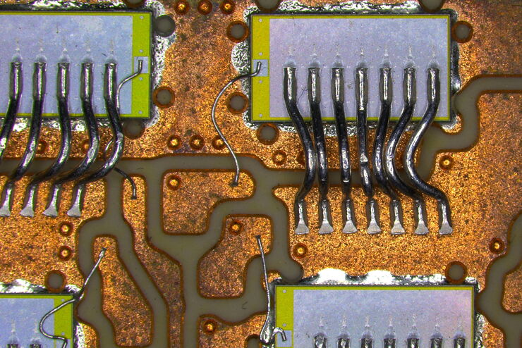
Inspection microscope image of a printed circuit board (PCB) taken with a ring light (RL) and near vertical illumination (NVI).

Microscope Illumination for Industrial Applications

Inspection microscope users can obtain information from this article which helps them choose the optimal microscope illumination or lighting system for inspection of parts or components.
Image of MDCK (Madin-Darby canine kidney) cells taken with phase contrast.

Phase Contrast and Microscopy

This article explains phase contrast, an optical microscopy technique, which reveals fine details of unstained, transparent specimens that are difficult to see with common brightfield illumination.

Immersion Objectives

How an immersion objective, which has a liquid medium between it and the specimen being observed, helps increase the numerical aperture and microscope resolution is explained in this article.
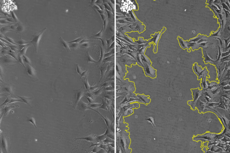
Phase-contrast image of a MDCK-cell culture and its respective confluency measured by the Mateo TL microscope.

How to Determine Cell Confluency with a Digital Microscope

This article shows how to measure cell confluency in an easy and consistent way with Mateo TL, increasing confidence in downstream experiments.
Intensity distribution (arbitrary color coding) of an image of two points where the distance between them corresponds to the Rayleigh criterion.

Microscope Resolution: Concepts, Factors and Calculation

This article explains in simple terms microscope resolution concepts, like the Airy disc, Abbe diffraction limit, Rayleigh criterion, and full width half max (FWHM). It also discusses the history.
[Translate to German:] Donor (D) and acceptor (A) molecule which participate in FRET (Förster resonance energy transfer).

Was ist FRET mit FLIM (FLIM-FRET)?

Der Beitrag erläutert die FLIM-FRET-Methode, die Resonanzenergietransfer und Fluoreszenz-Lebensdauer-Imaging zur Untersuchung von Protein-Protein Wechselwirkungen kombiniert.

Was ist FLIM - Fluorescence Lifetime Imaging Microscopy?

FLIM nutzt die Fluoreszenzlebensdauer, die misst, wie lange ein fluoreszierendes Molekül oder Fluorophor durchschnittlich in seinem angeregten Zustand bleibt, bevor es durch Aussenden eines…
Raw widefield and THUNDER image of transversal mouse adult fiber lens section. Courtesy N. Houssin, Plagemen lab, Ohio State University, Columbus, USA.

Studying Ocular Birth Defects

This article discusses how lens formation and ocular birth defects can be studied with sharp widefield microscopy images which are acquired rapidly. The mouse ocular lens is used as a model to study…

Cleanliness Analysis with a 2-Methods-in-1 Solution

In this article, it is examined how an overall efficient and cost-effective cleanliness analysis workflow can be achieved with a 2-methods-in-1 materials analysis solution, combining optical…
Automated Laser Microdissection for Proteome Analysis

Precise Spatial Proteomic Information in Tissues

Despite the availability of imaging-based and mass-spectrometry-based methods for spatial proteomics, a key challenge remains connecting images with single-cell-resolution protein abundance…
Image of murine-brain tissue showing a region removed with UV laser microdissection.

RNA Quality after Different Tissue Sample Preparation

The influence of sample preparation and ultraviolet (UV) laser microdissection (UV LMD) on the quality of RNA from murine-brain tissue cryo-sections is described in this article. To obtain good…
The various solutions from Leica Microsystems for cleanliness analysis.

Factors to Consider for a Cleanliness Analysis Solution

Choosing the right cleanliness analysis solution is important for optimal quality control. This article discusses the important factors that should be taken into account to find the solution that best…
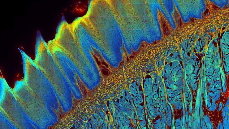
[Translate to German:] Transverse histological cut of a rabbit tongue. 50 Mpixels images (2326 µm x 1739 µm) in 14 x 18 tiles. Lifetime gives an additional contrast that allows to differentiate different structures in histological stainings.

Leitfaden zur Fluoreszenzlebensdauer-Imaging-Mikroskopie (FLIM)

The fluorescence lifetime is a measure of how long a fluorophore remains on average in its excited state before returning to the ground state by emitting a fluorescence photon.
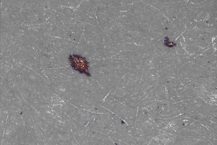
Type of contamination: spores

Cleanliness Analysis for Particulate Contamination

Devices, products, and their components fabricated in many industries can be quite sensitive to contamination and, as a result, have stringent requirements for technical cleanliness. Measurement…
[Translate to German:] Particles and fibers on a filter which will be counted and analyzed for cleanliness

Effiziente Partikelzählung und -analyse

Dieser Bericht befasst sich mit der Partikelzählung und -analyse unter Verwendung der optischen Mikroskopie bei der technischen Sauberkeitsanalyse von Teilen und Komponenten. Die Partikelzählung und…
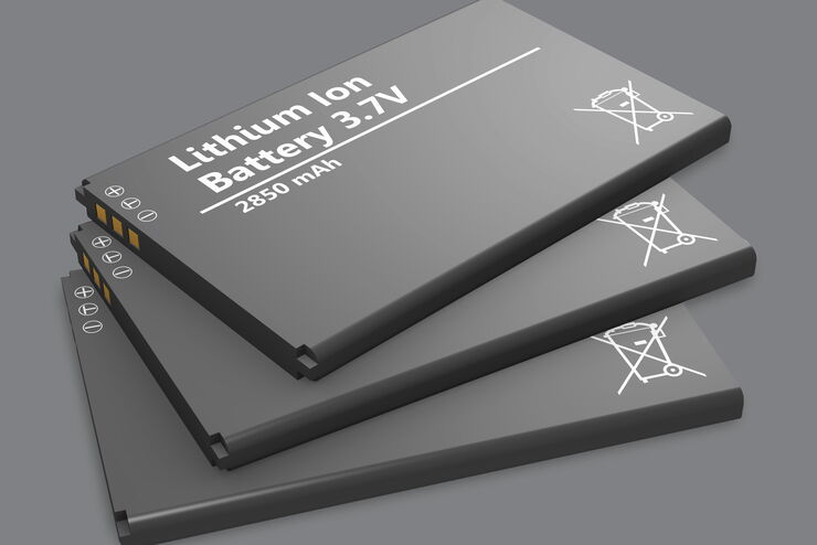
A stack of lithium-ion batteries

Quality Control Under the Microscope

Fast-rising demand for electric vehicles is one of the market’s main drivers, but there are other hotspots of growth, including the rise in renewable energy installations, such as photovoltaic panels,…
When particulate contamination is present in lubricating fluids or oils, it can cause damage to parts or components leading to malfunctions.

Hydraulics in the Automotive and Aerospace Industries

Cleanliness standards relating to lubricants, hydraulic fluids, and oils, e.g., ISO 4406 and DIN 51455 are discussed in this article. Cleanliness plays a central role in the automotive and…
Scroll to top