Sciences de la vie
Sciences de la vie
C'est ici que vous pourrez développer vos connaissances, vos capacités de recherche et les applications pratiques de la microscopie dans divers domaines scientifiques. Apprenez à obtenir une visualisation précise, à interpréter les images et à faire progresser la recherche. Trouvez des informations pertinentes sur la microscopie avancée, les techniques d'imagerie, la préparation des échantillons et l'analyse des images. Les sujets abordés comprennent la biologie cellulaire, les neurosciences et la recherche sur le cancer, en mettant l'accent sur les applications et les innovations de pointe.
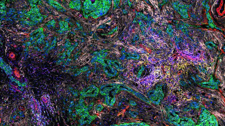
AI-Powered Hi-Plex Spatial Analysis Tools for Breast Cancer Research
Breast cancer (BC) is the leading cause of cancer-related deaths in women. Investigating the tumor microenvironment (TME) is crucial to elucidate the mechanisms of tumor progression. Systematic…
Neurosciences
Vous souhaitez une meilleure compréhension des maladies neurodégénératives ou vous étudiez le fonctionnement du système nerveux ? Découvrez comment vous pouvez faire des percées importantes grâce aux…
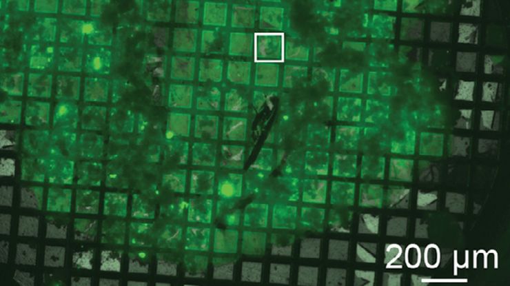
The “Waffle Method”: High-Pressure Freeze Complex Samples
This article describes the advantages of a special high pressure freezing method, the so-called “Waffle Method”. Learn how the “Waffle Method” uses EM grids as spacers for high-pressure freezing,…
Mastering Polymer Sectioning with Helmut Gnaegi
When it comes to ultramicrotomy, few names carry the weight of Helmut Gnaegi. As co-founder of Diatome, a global leader in diamond knife technology, Helmut has spent decades refining the art and…
A Guide to Zebrafish Research - See More Details at a Glance
To obtain optimal results while doing zebrafish research, especially during screening, sorting, handling, and imaging, seeing the fine details and structures is important. They help researchers make…
How to Streamline High-Plex Imaging for 3D Spatial Omics Advances
In this webinar, Dr. Julia Roberti and Dr. Luis Alvarez from Leica Microsystems introduce SpectraPlex, a new functionality integrated into the STELLARIS confocal platform for high-plex 3D spatial…
Improving Zebrafish-Embryo Screening with Fast, High-Contrast Imaging
Discover from this article how screening of transgenic zebrafish embryos is boosted with high-speed, high-contrast imaging using the DM6 B microscope, ensuring accurate targeting for developmental…
Transforming Research with Spatial Proteomics Workflows
Spatial Proteomics, Nature Methods 2024 Method of the Year, is driving research advancements in cancer, immunology, and beyond. By combining positional data with high throughput imaging of proteins in…
How Fluorescence Guides Sectioning of Resin-embedded EM Samples
Electron microscopes, including transmission electron microscopes (TEM) and scanning electron microscopes (SEM), are widely utilized to gain detailed structural information about biological samples or…


