
Scienze della vita
Scienze della vita
Questo è il posto giusto per ampliare le vostre conoscenze, le capacità di ricerca e le applicazioni pratiche della microscopia in vari campi scientifici. Imparate come ottenere una visualizzazione precisa, l'interpretazione delle immagini e i progressi della ricerca. Troverete informazioni approfondite sulla microscopia avanzata, sulle tecniche di imaging, sulla preparazione dei campioni e sull'analisi delle immagini. Gli argomenti trattati comprendono la biologia cellulare, le neuroscienze e la ricerca sul cancro, con particolare attenzione alle applicazioni e alle innovazioni più avanzate.
 stereo microscope for a task like surgery.")
Rodent and Small-Animal Surgery
Learn how you can perform rodent (mouse, rat, hamster) and small-animal surgery efficiently with a microscope for developmental biology and medical research applications by reading this article.

Imaging and Analyzing Zebrafish, Medaka, and Xenopus
Discover how to image and analyze zebrafish, medaka, and Xenopus frog model organisms efficiently with a microscope for developmental biology applications from this article.

Investigating Fruit Flies (Drosophila melanogaster)
Learn how to image and investigate Drosophila fruit fly model organisms efficiently with a microscope for developmental biology applications from this article.

Studying Caenorhabditis elegans (C. elegans)
Find out how you can image and study C. elegans roundworm model organisms efficiently with a microscope for developmental biology applications from this article.
")
Epoxy Resin Embedding of Animal and Human Tissues for Pathological Diagnosis and Research
Application Note for Leica EM AMW - The tissues were fixed in the modified Karnovsky fixative generally by immersion overnight (at minimum 4h at room temperature). Then pieces of approx. 1mm3 were cut…

Brief Introduction to Freeze Substitution
Freeze-substitution is a process of dehydration, performed at temperatures low enough to avoid the formation of ice crystals and to circumvent the damaging effects observed after ambient-temperature…
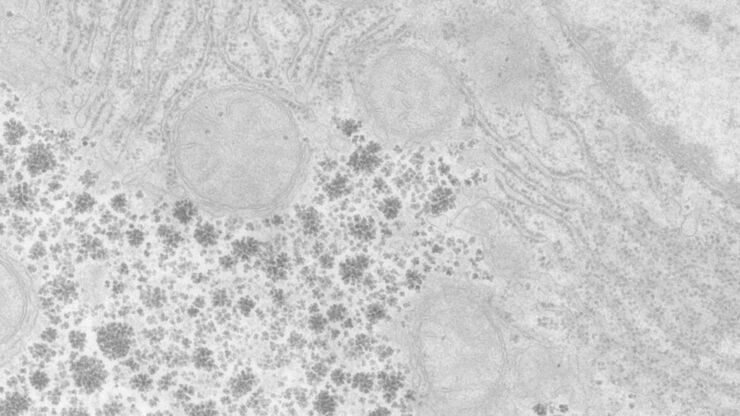
Perusing Alternatives for Automated Staining of TEM Thin Sections
Contrast in transmission electron microscopy (TEM) is mainly produced by electron scattering at the specimen: Structures that strongly scatter electrons are referred to as electron dense and appear as…
An Introduction to CARS Microscopy
CARS overcomes the drawbacks of conventional staining methods by the intrinsic characteristics of the method. CARS does not require labeling because it is highly specific to molecular compounds which…
")
Mosaic Images
Confocal laser scanning microscopes are widely used to create highly resolved 3D images of cells, subcellular structures and even single molecules. Still, an increasing number of scientists are…
