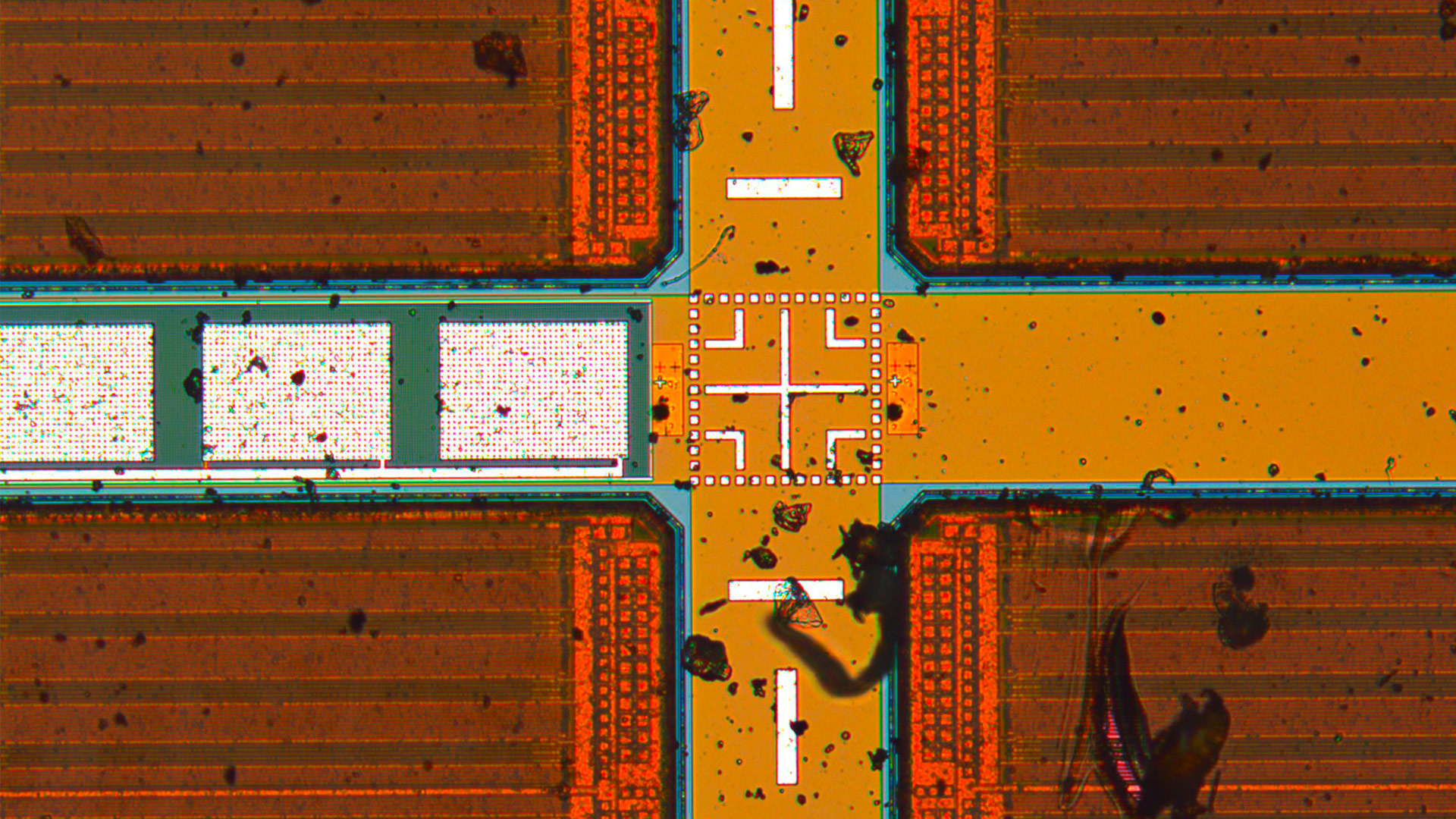

Meet Mica
The world’s first Microhub
Mica enables microscopy access for all, removes the constraints of traditional four-color fluorescence imaging and radically simplifies workflows.
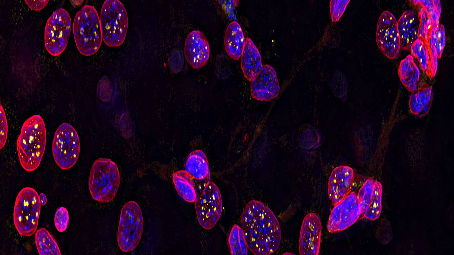
Cell Biology Research
Imaging solutions offered by Leica Microsystems are designed to maximize your cell biology research.
Follow us on Instagram
Frequently Asked Questions Light Microscopes
Leica microscopes are modular and shipped in the configuration that best fits your stated needs or application. In case your needs change later, you can always upgrade your workstation by adding available accessories.
Leica Microsystems offers the free Store & Recall software (available with the LAS X software platform for industry) which allows you to customize the microscope functions, adapting them to the requirements and needs of each individual user. The software also allows you to restore all system settings saved with the acquired image.
All our encoded microscope solutions offer calibration and image comparison. Furthermore, Leica Microsystems offers the free Store & Recall software which allows you to restore all system settings saved with the acquired image.
No, you don’t. By installing a FLEXACAM C1 camera, you can directly save the images on an IT network server or USB medium. You can also send the images via e-mail over your network without the need for a PC.
With the FLEXACAM C1 camera you can directly save images on an IT network server or USB medium. You can also send the images via e-mail without having to use a computer.
There are a lot of accessories. Please get in contact with your local Leica sales representative.
There are a lot of ergonomic accessories for Leica light microscopes. Please contact your local Leica sales representative for more details or visit: Ergonomic Accessories for Stereo Microscopes
LAS X software only runs with Windows, but for a MAC we have a dedicated software called Leica Acquire that you could download for free from the Apple store: https://apps.apple.com/it/app/leica-acquire/id733706983?mt=12
However, there is no software for Linux.
Yes, use of a 3rd party software is possible: https://www.splashtop.com/classroom
We have adapters for all C-mount compatible cameras.
The free AirLab software, compatible with iOS or ANDROID, allows the user to immediately share images, videos, and comments.
A compound microscope uses optics to produce a magnified image of a sample so that with details of it can be observed that are undetectable with the naked eye. The most basic optics of a compound microscope has at least 2 lenses: i) an objective placed nearby the sample which creates a magnified, real image of it and ii) eyepieces or oculars which are used to view the real image of the sample. A human who looks through the eyepieces sees the sample as a virtual image on his/her retina. For more information, please refer to the Science Lab article: Optical Microscopes – Some Basics
The maximum useful magnification value achieved with any type of light microscope depends on its ultimate resolving power or maximal resolution. The resolution depends on the microscope objective lens numerical aperture (NA). At low magnification values, the NA is small leading to a low resolution. At high magnification values, the NA is high yielding a high resolution. However, because the NA has a finite maximum value, approximately 1.3, the “useful” range of magnification is limited to about 1,800x for conventional light microscopes. Magnification beyond the useful range, which can occur whenever digital microscope cameras display images on large monitors, is called "empty” magnification. In this non-useful magnification range, the sample structures appear larger, but no additional details are resolved. For more information, please refer to the Science Lab articles: Beware of "Empty" Magnification, What Does 30,000:1 Magnification Really Mean?