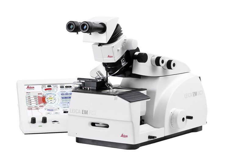
Consistent high-quality sections at room and cryo temperatures
Ultramicrotome Leica EM UC7
Read our latest articles

Exploring the Structure and Life Cycle of Viruses
The SARS-CoV-2 outbreak started in late December 2019 and has since reached a global pandemic, leading to a worldwide battle against COVID-19. The ever-evolving electron microscopy methods offer a…
Read article

Investigating Synapses in Brain Slices with Enhanced Functional Electron Microscopy
A fundamental question of neuroscience is: what is the relationship between structural and functional properties of synapses? Over the last few decades, electrophysiology has shed light on synaptic…
Read article

Workflows and Instrumentation for Cryo-electron Microscopy
Cryo-electron microscopy is an increasingly popular modality to study the structures of macromolecular complexes and has enabled numerous new insights in cell biology. In recent years, cryo-electron…
Read article

Upright Fluorescence Microscopy for Virus Replication Studies
The severe acute respiratory syndrome coronavirus 2 (SARS-CoV-2) emerged in late 2019 and spread over the world quickly. Due to its dramatic impact, researchers are interested in the nature of the…
Read article

Microscopy in Virology
The coronavirus SARS-CoV-2, causing the Covid-19 disease effects our world in all aspects. Research to find immunization and treatment methods, in other words to fight this virus, gained highest…
Read article

Workflow Solutions for Industrial Research
This brochure presents and explains appropriate workflow solutions for the most frequently required sample preparation methods for material science samples.
Read article

Medicinal Use of Qianhu Plant Roots
This study focused on tissue-specific metabolite profiling and comparison of various parts of bolting and unbolting Peucedanum praeruptorum Dunn (Qianhu in Chinese). The qualitative and quantitative…
Read article

Ultrastructural Preservation and Improved Visualization of Membranes in Primary Bovine Chromaffin Cells
Intracellular dynamic events, such as protein trafficking, recycling, and degradation as well as signal transduction, to name a few, can be studied using live-cell imaging techniques. However,…
Read article

Introduction to Ultramicrotomy
When studying samples, to visualize their fine structure with nanometer scale resolution, most often electron microscopy is used. There are 2 types: scanning electron microscopy (SEM) which images the…
Read article

Bridging Structure and Dynamics at the Nanoscale through Optogenetics and Electrical Stimulation
Nanoscale ultrastructural information is typically obtained by means of static imaging of a fixed and processed specimen. However, this is only a snapshot of one moment within a dynamic system in…
Read article

Superior Ultrastructural Preservation and Structural Contrast in Drosophila Tissue
Optimal structural preservation of tissue can only be obtained by high pressure freezing. However, preparing the samples in optimal conditions is challenging. This article explains in detail how to…
Read article

Variety of RNAs in Peripheral Blood Cells, Plasma, and Plasma Fractions
Human peripheral blood contains RNA in cells and in extracellular membrane vesicles, microvesicles and exosomes, as well as in cell-free ribonucleoproteins. Circulating mRNAs and noncoding RNAs, being…
Read article

Cryo-Electron Microscopy of Vitreous Sections (CEMOVIS) of Yeast
Application Note for Leica EM HPM100 - The sections are of yeast frozen with a Leica HPM100 high pressure freezer in the copper tube system, the cell paste was mixed with a pH 6.5 MES/dextran buffer…
Read article

Immuno - Electron Microscopy of High Pressure Frozen and Freeze Substituted Mouse Heart
Application Note for Leica EM AFS2 - Mouse heart tissue from wild-type (WT) and αT -catenin KO (KO) mice was excised, immersed in 20% (w/v) BSA and frozen immediately in a high-pressure freezer Leica…
Read article

Cell Envelope Structure of a Gram-negative Thermotolerant y-Proteobacterium Acidithiobacillus sp., Strain HV2/2 and its Interaction with Pyrite
Application Note for Leica EM AFS2 - Cells of the the moderately thermophilic Acidithiobacillus sp.strain HV2/2, were centrifuged at 20,0009xg and then cryo-immobilized by high-pressure freezing on…
Read article

Symmetric Synapse - Clathrin Coated Endocytosis Pit in the Postsynaptic Dendrite
Application Note for Leica EM ICE - WT hippocampal neurons were plated at a density of 80,000 cells/cm2 on 6 mm sapphire disks for 14 days. Sample were frozen using a high-pressure freezer (Leica EM…
Read article

Electron Microscopy of High Pressure Frozen and Freeze Substituted Arabidopsis Thaliana Root Tips Cells
Application Note for Leica EM AFS2 - Arabidopsis thaliana roots (mutant PIN1pro:PIN1-GFP;bex5-1) were excised, immersed in 20% (w/v) BSA and frozen immediately in a high-pressure freezer. Freeze…
Read article

Electron Microscopy Sample Preparation: “The Future is Cold, Dynamic and Hybrid”
In 2014, the renowned Electron Microscopy for Materials Science (EMAT) research lab at the University Antwerp, Belgium, and Leica Microsystems started a fruitful collaboration to establish a Leica…
Read article

Transmission Electron Microscopy Sample Preparation Protocols for the Ultrastructural Study of Cysts of Free-living Protozoa
Cysts of free-living protozoa have an impact on the ecology and epidemiology of bacteria because they may act as a transmission vector or shelter the bacteria against hash environmental conditions.…
Read article

Correlative In-Resin Super-Resolution and Electron Microscopy Using Standard Fluorescent Proteins
We introduce a method for correlative in-resin super-resolution fluorescence and electron microscopy (EM) of biological structures in mammalian culture cells. Cryo-fixed resin embedded samples offer…
Read article

Influence of Tissue and Plant Species in the Trafficking of a Recombinant Protein in Plant Cells
The development of recombinant DNA technology has allowed the use of plants for the production of biopharmaceuticals. In contrast to other production platforms, plants are unexpensive, easy to scale…
Read article

Protein Trafficking in Cereal Seeds
Seeds accumulate proteins and starch which will be broken down and mobilized upon germination. Among seeds, cereals constitute an example of a highly specialized storage tissue that constitutes up to…
Read article

Perusing Alternatives for Automated Staining of TEM Thin Sections
Contrast in transmission electron microscopy (TEM) is mainly produced by electron scattering at the specimen: Structures that strongly scatter electrons are referred to as electron dense and appear as…
Read article

Atomic Force Microscopy Study of a Stretched Impact Copolymer
Atomic force microscopy (AFM) is a powerful tool in the suite of nanoscale characterization techniques that provides a variety of information including topography, mechanical properties, and…
Read article

Focussed Ion Beam Milling and Scanning Electron Microscopy of Brain Tissue
This protocol describes how biological samples, like brain tissue, can be imaged in three dimensions using the focussed ion beam/scanning electron microscope (FIB/SEM). The samples are fixed with…
Read article

Advanced Correlative Light/Electron Microscopy: Current Methods and New Developments Using Tokuyasu Cryosections
Microscopy is an essential tool for analysis of cellular structures and function. With the advent of new fluorescent probes and super-resolution light microscopy techniques, the study of dynamic…
Read article
Fields of Application

Sample Preparation for Materials Science
Sample preparation is key for observing the finest textures and details on the sample surface in high resolution. The right workflow and tools for the pre-preparation will lead to an excellently…
Read article

Cell Culture
Growing cells under lab conditions is the base for scientists working in fields of cell or developmental biology, cancer research, or any kind of life science and pharma research. Find out how Leica…
Read article

Zebrafish Research
For the best result during screening, sorting, manipulation, and imaging you need to see details and structures to make the right decisions for your next steps in research.
Known for outstanding…
Read article

Fluorescence Microscopy Solutions
Find out how fluorescence microscopes from Leica Microsystems support your research. Fluorescence is one of the most commonly used physical phenomena in biological and analytical microscopy for its…
Read article

Life Science Education
Microscopes for life science education from Leica Microsystems are high-performing, durable imaging systems meet your classroom needs at affordable prices.
Read article