Peter Laskey , Dr.

Peter Laskey

Pete Laskey studied Biological and Medicinal Chemistry, he then competed a masters in Pharmacology and a PhD in Virology. During his PhD he studied the interaction between the human papilloma virus E1^E4 protein and cell cytoskeleton specializing in live cell imaging. He went on to work as an imaging specialist at the National Institute of Medical Research before leaving to spend several years traveling the world. On returning to Europe he spent several years at Andor focusing on sCMOS camera technology before joining Leica as the Product Manager for the GSD 3D super resolution system in 2013.

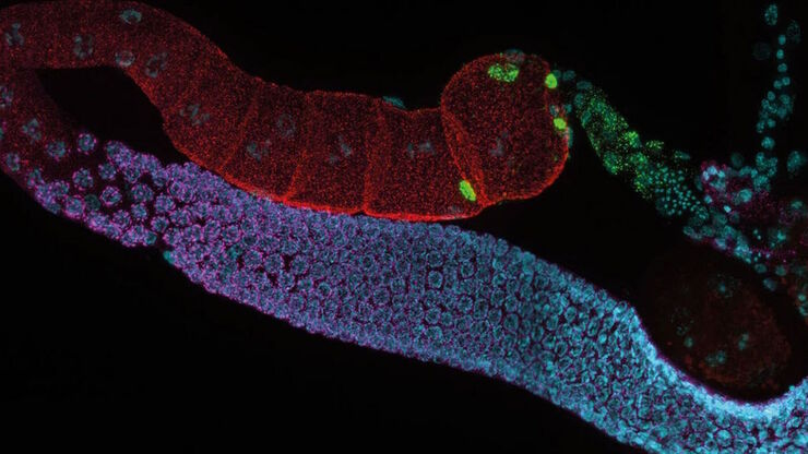
C. elegans adult hermaphrodite gonades acquired using THUNDER Imager. Staining: blue - DAPI (nucleus), green - SP56 (sperm), red - RME-2 (oocyte), magenta - PGL-1 (RNA + protein granules). Image courtesy of Prof. Dr. Christian Eckmann, Martin Luther University, Halle, Germany.

Life Science Research: Which Microscope Camera is Right for You?

Deciding which microscope camera best fits your experimental needs can be daunting. This guide presents the key factors to consider when selecting the right camera for your life science research.
Staining is crucial to revealing details in the specimens.

H&E Staining in Microscopy

If we consider the role of microscopy in pathologists’ daily routines, we often think of the diagnosis. While microscopes indeed play a crucial role at this stage of the pathology lab workflow, they…
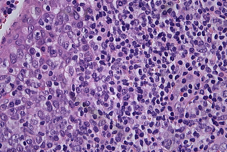
Histopathological sample, 40x magnification

Clinical Microscopy: Considerations on Camera Selection

The need for images in pathology laboratories has significantly increased over the past few years, be it in histopathology, cytology, hematology, clinical microbiology, or other applications. They…
Microscope Camera K3M

Introduction to Digital Camera Technology

A significant majority of modern optical microscopy techniques require the use of a digital camera. By working with digital devices researchers can observe specimens on a screen in real time or…
Scroll to top