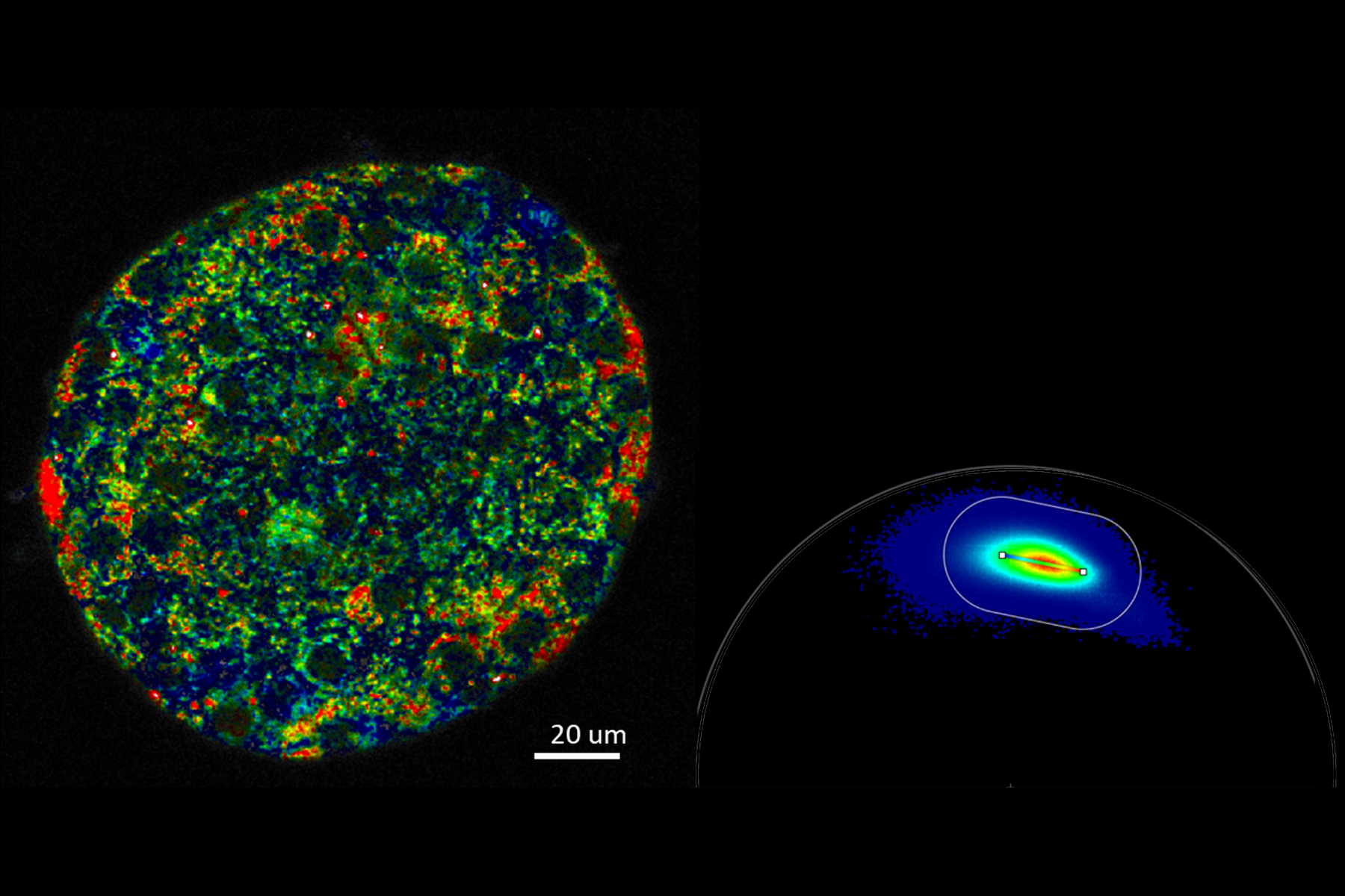
The maintenance of normal blood glucose levels is defective in diabetes caused by dysfunction of the alpha and beta cells in islets. To understand these regularities, we used multiphoton phasor-FLIM NADH autofluorescence imaging to detect metabolic changes of living islet cells before and after glucose stimulation. Multiphoton phasor FLIM NADH autofluorescence imaging provides a straightforward detection and analysis to monitor metabolic states in living organisms.
Meanwhile, it provides the high spatial resolution to monitor alpha cells and beta cells separately in a time lapse. We observed increased oxidative phosphorylation in beta cells and suppressed oxidative phosphorylation in alpha cells after glucose stimulation in healthy islets, which is not seen in islets with type 2 diabetes. This proved that phasor FLIM could be a powerful tool to monitor cell metabolism and drug discovery in diabetes.
Image: Metabolic imaging of mouse pancreatic islet.


 in 14 x 18 tiles. Lifetime gives an additional contrast that allows to differentiate different structures in histological stainings.")
