Access to Webinar
Access the free webinar-on demand on BitesizeBio.com:
Key Learnings
We present the SP8 FALCON from Leica Microsystems, a new high-end confocal instrument with built-in very fast FLIM capabilities. The instrument applies a novel technology to records photon arrival times at up to 80 MHz (160 MHz if two detectors are used) using the built-in spectral HyD detectors. To arrive at such high counts rates, the instrument uses real-time 10 GHz sampling and analysis algorithms that render it largely immune to pulse pile-up. Falcon FLIM has been fully integrated in the LAS-X software, allowing convenient acquisition of FLIM time-lapses, stitched FLIM overview images, FLIM XYZ-stacks and spectral FLIM stacks, among others, as well as combinations thereof.
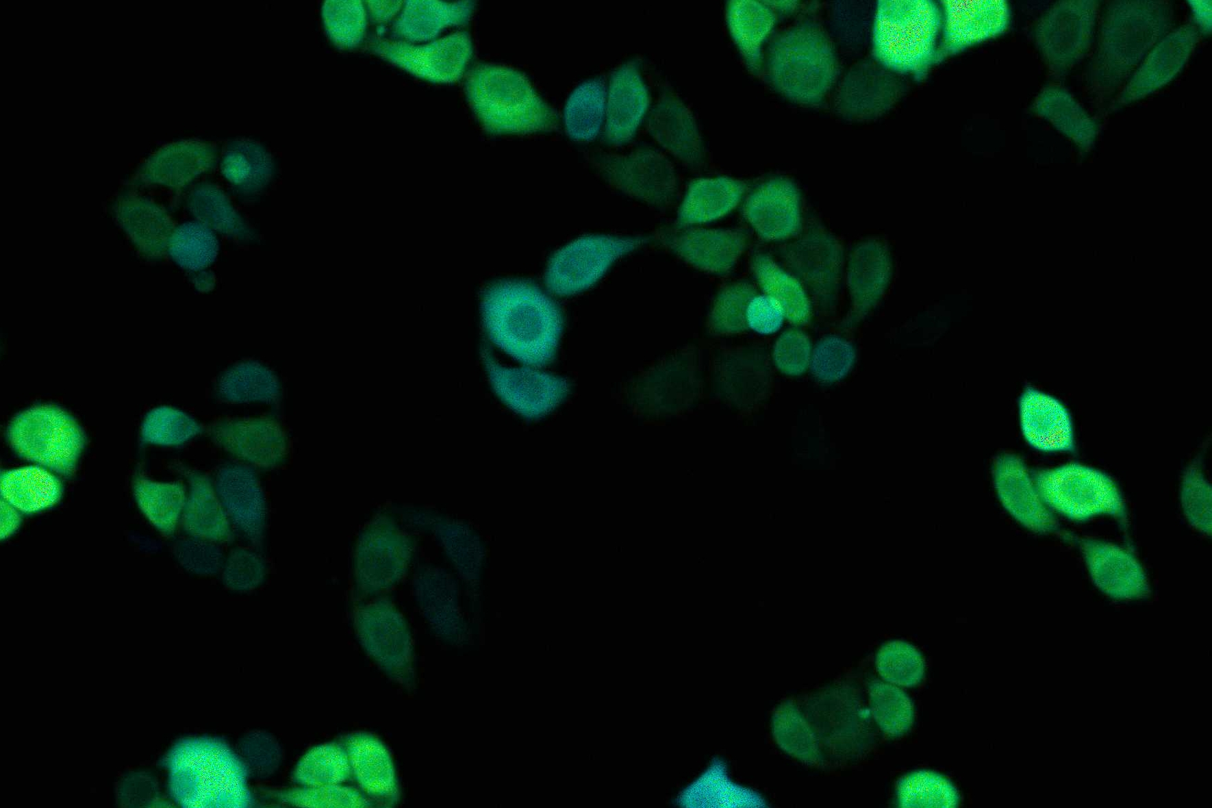
We scrutinized the performance of the SP8 FALCON and applied it to record agonist-induced changes in concentration of second messengers such as cAMP and Ca2+ with sub-micrometer precision at high speed- on the order of several (512×512) frames per second (FPS). To detect cAMP by FLIM, we used our newest generation of EPAC-based dedicated FLIM sensors, which use mTurquoise2 as a donor and a tandem of two monomeric dark Venus proteins as acceptors. Reducing the image format to 128×128 pixels, we acquired good quality FLIM time-lapses at 25 FPS (83 FPS using resonant scanning), allowing detection of even very fast signaling events, including e.g. the activation of G-proteins and Ca2+ sparks. The large numbers of detected photons reduce pixel-to-pixel variability in the calculated lifetimes. Finally we demonstrate that the SP8-FALCON is ideal for fast FLIM screening applications in both fixed- and live-cell formats.


