STELLARIS FALCON
Konfokalmikroskope
Produkte
Startseite
Leica Microsystems
STELLARIS FALCON Lebenszeit-Messung im Nu
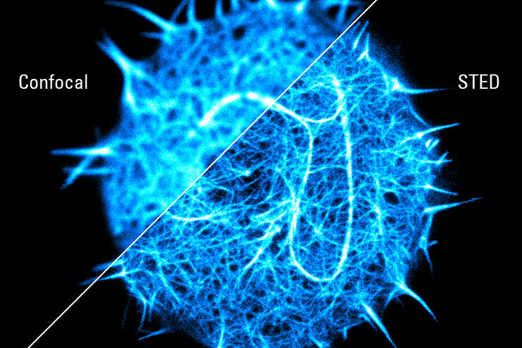
Der Kontrast ist deutlich
Lesen Sie unsere neuesten Artikel
 and acceptor (A) molecule which participate in FRET (Förster resonance energy transfer).")
Was ist FRET mit FLIM (FLIM-FRET)?
Der Beitrag erläutert die FLIM-FRET-Methode, die Resonanzenergietransfer und Fluoreszenz-Lebensdauer-Imaging zur Untersuchung von Protein-Protein Wechselwirkungen kombiniert.

Researchers Insights: Microscopy in Cancer Research
Discover how imaging techniques are driving cancer research forward. In this issue, we present comprehensive multimodal studies using microscopy, as well as new directions in intraoperative cancer…
, Tropomyosin (cardiomyocytes, red) and GFP (primordial cardiac layer, green).")
A Guide to Fluorescence Microscopy
Fluorescence microscopy uses the ability of fluorophores, dyes, or fluorescent proteins to emit light of a specific wavelength after being excited with light of a shorter wavelength. Biomolecules can…
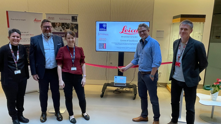
Die Micron Bioimaging Facility an der University of Oxford
Erfahren Sie, wie Sie am Department of Biochemistry der University of Oxford Zugang zu modernster Mikroskopietechnologie sowie gezielter Schulung und fachkundiger Unterstützung erhalten.
. Courtesy: Thomas Mathivet, PhD")
Windows on Neurovascular Pathologies
Discover how innate immunity can sustain deleterious effects following neurovascular pathologies and the technological developments enabling longitudinal studies into these events.

Live-Cell Fluorescence Lifetime Multiplexing Using Organic Fluorophores
On-demand video: Imaging more subcellular targets by using fluorescence lifetime multiplexing combined with spectrally resolved detection.

Visualizing Protein-Protein Interactions by Non-Fitting and Easy FRET-FLIM Approaches
The Webinar with Dr. Sergi Padilla-Parra is about visualizing protein-protein interaction. He gives insight into non-fitting and easy FRET-FLIM approaches.
,由14 × 18个图块拼接而成。荧光寿命提供了额外的对比度,使组织染色中的不同结构得以区分。")
Leitfaden zur Fluoreszenzlebensdauer-Imaging-Mikroskopie (FLIM)
Die Fluoreszenzlebensdauer ist ein Maß dafür, wie lange ein Fluorophor im Durchschnitt in seinem angeregten Zustand verbleibt, bevor er durch Aussendung eines Fluoreszenzphotons in den Grundzustand…

Find Relevant Specimen Details from Overviews
Switch from searching image by image to seeing the full overview of samples quickly and identifying the important specimen details instantly with confocal microscopy. Use that knowledge to set up…

Fluorescence Lifetime-based Imaging Gallery
Confocal microscopy relies on the effective excitation of fluorescence probes and the efficient collection of photons emitted from the fluorescence process. One aspect of fluorescence is the emission…

How to Quantify Changes in the Metabolic Status of Single Cells
Metabolic imaging based on fluorescence lifetime provides insights into the metabolic dynamics of cells, but its use has been limited as expertise in advanced microscopy techniques was needed.
Now,…

How FLIM Microscopy Helps to Detect Microplastic Pollution
The use of autofluorescence in biological samples is a widely used method to gain detailed knowledge about systems or organisms. This property is not only found in biological systems, but also…

Mit LIGHTNING das Maximum an Informationen aus Ihrer Probe erhalten
LIGHTNING ist ein adaptiver Prozess zur Extraktion von Bildinformationen, bei dem vollautomatisch anderweitig nicht sichtbare Strukturen und feine Details sichtbar gemacht werden. Im Gegensatz zu…

Virologie
Liegt Ihr Forschungsschwerpunkt auf Virusinfektionen und -krankheiten? Erfahren Sie, wie Sie mit Lösungen für Bildgebung und Probenvorbereitung von Leica Microsystems mehr Erkenntnisse in der…

Microscopy in Virology
The coronavirus SARS-CoV-2, causing the Covid-19 disease effects our world in all aspects. Research to find immunization and treatment methods, in other words to fight this virus, gained highest…

TauSense Technology Imaging Tools
Leica Microsystems’ TauSense technology is a set of imaging modes based on fluorescence lifetime. Found at the core of the STELLARIS confocal platform, it will revolutionize your imaging experiments.…
Super-resolved STED spectroscopy
Molecular interactions are key in cellular signalling. They are often ruled or rendered by the mobility of the involved molecules.

Förster Resonance Energy Transfer (FRET)
The Förster Resonance Energy Transfer (FRET) phenomenon offers techniques that allow studies of interactions in dimensions below the optical resolution limit. FRET describes the transfer of the energy…
Anwendungsbereiche
Erweiterte Gewebebildgebung und -analyse
Gewinnen Sie mit den erweiterten Bildgebenden Verfahren von Leica Microsystems Einblicke in die Gewebestruktur und -funktion und verbessern Sie Ihr Verständnis der räumlichen Biologie und der…
