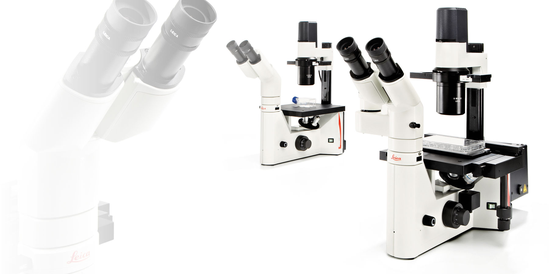
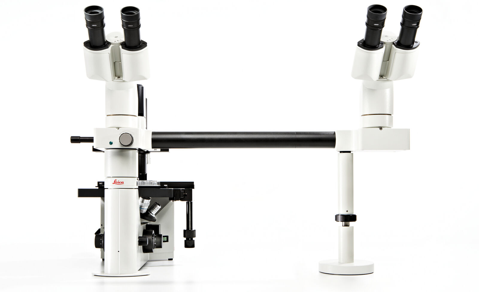
DM IL LED Inverted Laboratory Microscope
Ideal for cell culture, micromanipulation, documentation of immunostained specimens, and routine live cell examinations. Monitor your specimen the way you need: Phase contrast, modulation contrast and fluorescence are just one fingertip away.
Robust stability, plenty of space to work with tools, long working distances to accommodate large culture flasks and a stable illumination without heat make work at the microscope easy and convenient.
For special diagnostics requirements, the microscope is certified for in-vitro-diagnostics (IVD) including in-vitro-fertilization (IVF).
Show popular configurations
Explore and buy pre-configured microscopy solutions in our online shop. Enjoy a seamless online shopping experience.
Contrasting methods
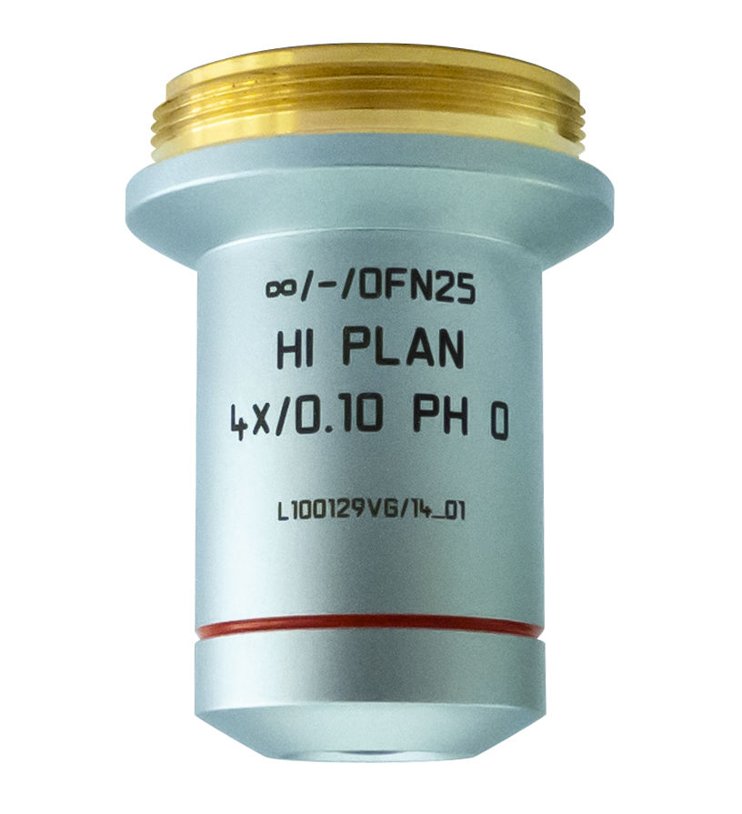
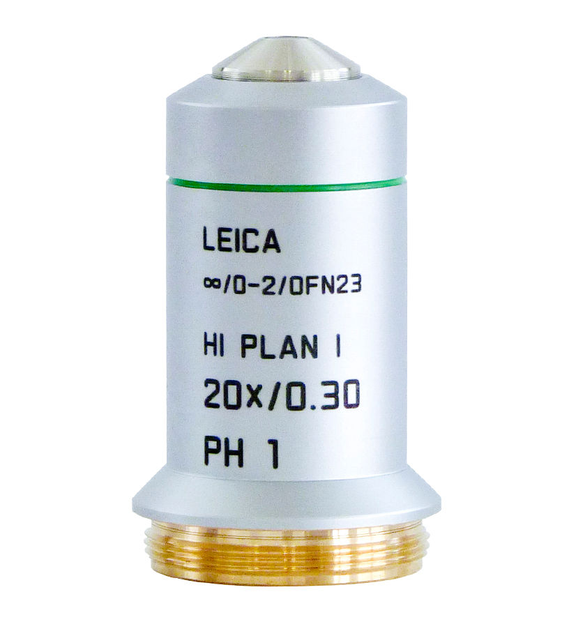
In addition to Phase Contrast and Brightfield illumination, the DM IL features unique Integrated Modulation Contrast (IMC) from Leica without the need for special objectives.
Available for 10x, 20x, 32x, and 40x magnification.

Cell culture
With a long working distance the standard DM IL microscope allows you to conveniently monitor large cell culture flasks.
If your cell production reaches the next level, DM IL Cell Factory is the right choice. With a transmitted light arm of 480 mm, large multilayer vessels with up to 10 chambers can be accommodated, enabling imaging of lower layers - including fluorescence.
Please contact your local sales representative for further information.

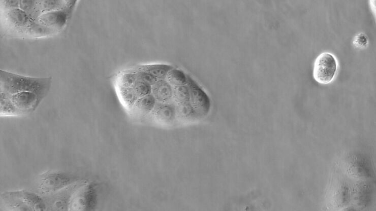
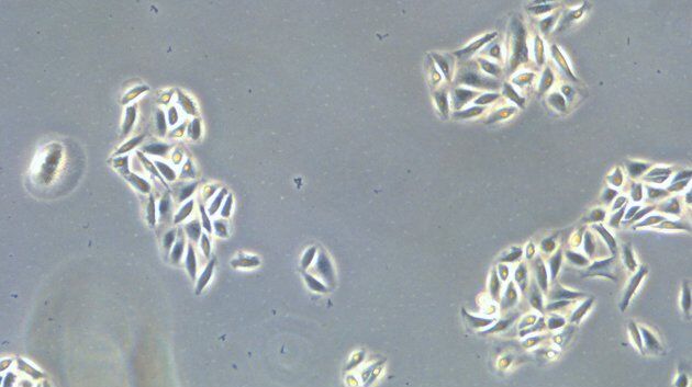
Live cell imaging
When observing living cells under a microscope, it’s essential to maintain optimal conditions for the organisms. Leica Microsystems offers a wide range of accessories for any application, so the environmental conditions can be controlled easily during one experiment.
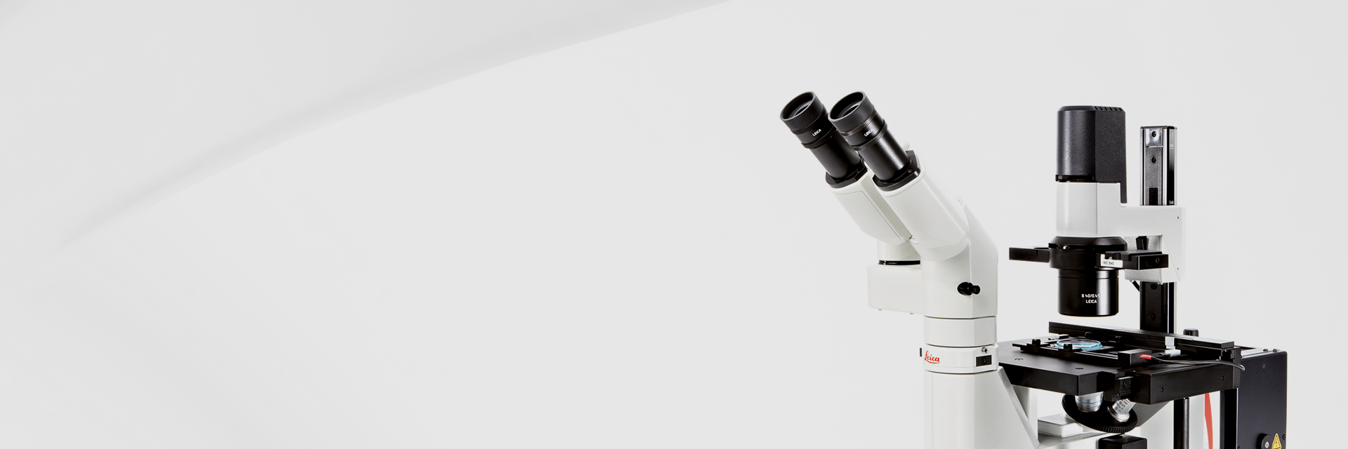
Relaxed and comfortable
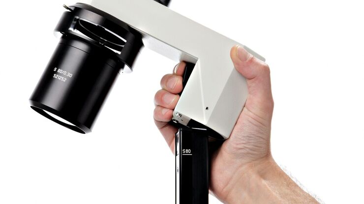
The ergonomic design of controls such as the focus dial, brightness controller, condenser height adjustment, objective nosepiece, and XY stage adjustment allows the user to comfortably work and manipulate their sample.
The microscope can even adapt to different users with height-adjustable stages and ergo tubes with variable height.

Product life of 50,000 hours
High-intensity, high-contrast 5W LED illumination with a product life of 50,000 hours provides a constant color temperature. This makes documentation of your results remarkably easy and reliable. The brightness of the LED automatically adjusts to the contrast method which reduces the time needed for manual interactions.


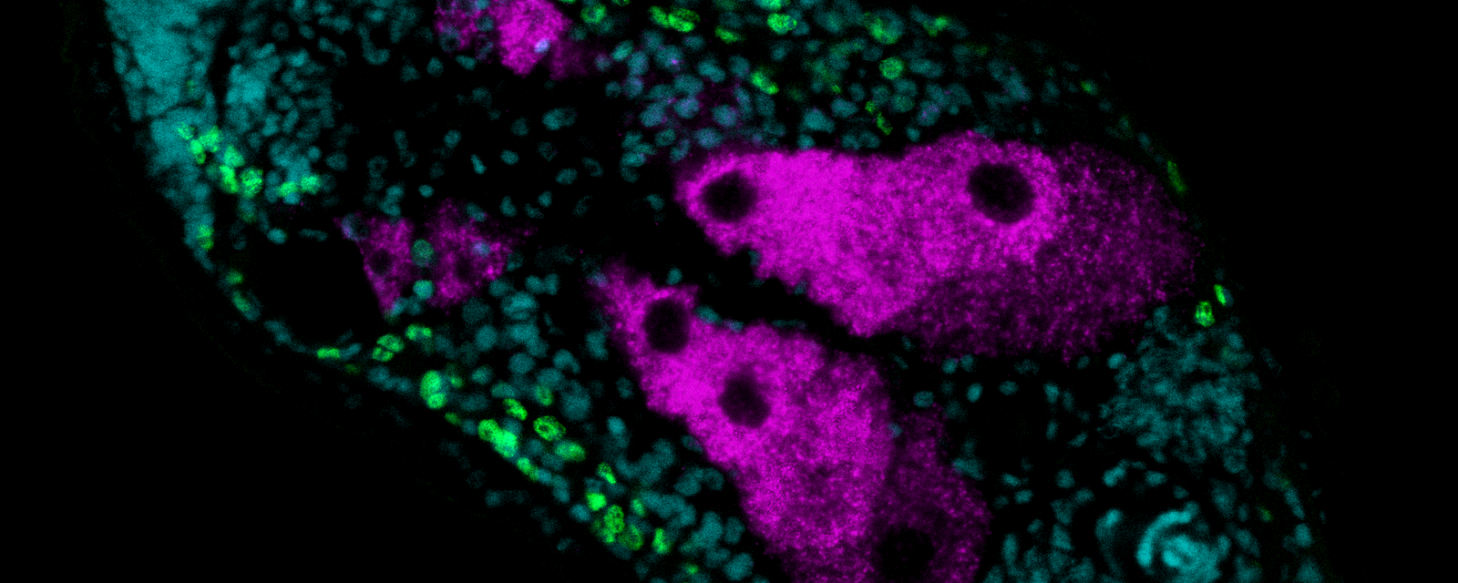
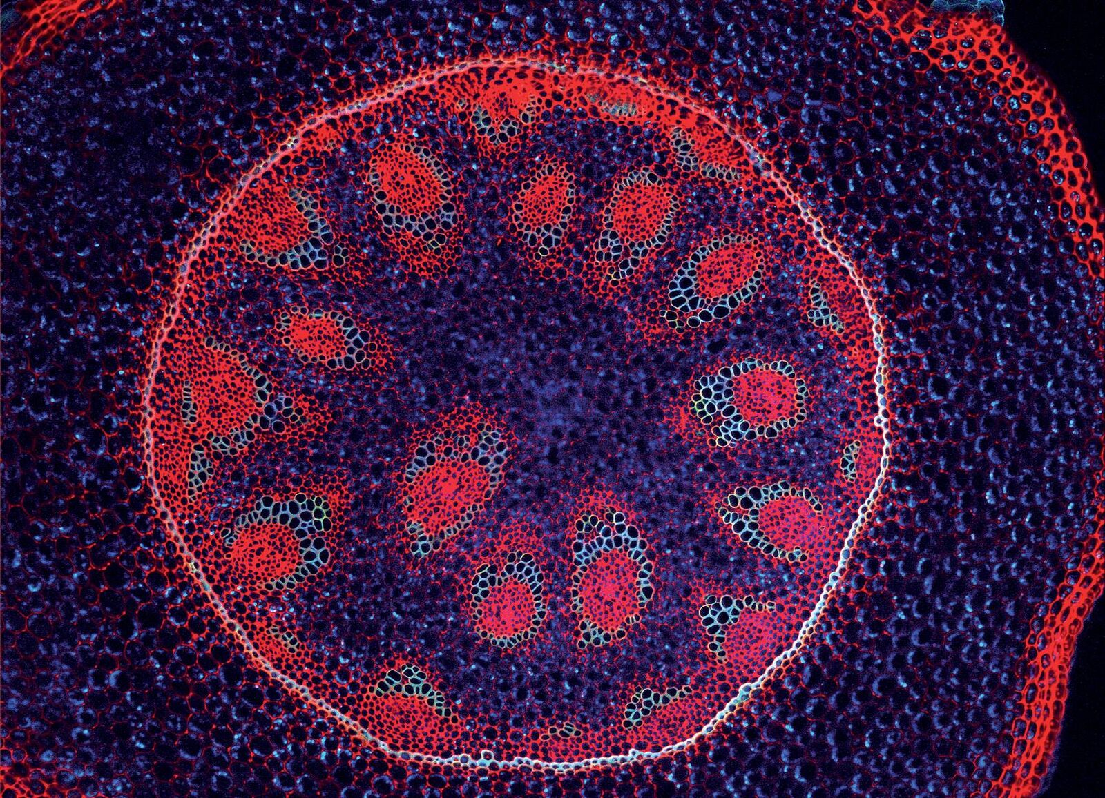
Fluorescence
Equipped with a 3 position fluorescence slider, the DMIL is capable of capturing brilliant fluorescence images. Confidently overlay multiple fluorescence images due to zero pixel shift technology.
Easily add another slider to image more fluorochromes.
Documentation
A broad portfolio of digital cameras for brightfield and fluorescence documentation complements the DM IL LED imaging system.
Together with powerful LAS X software, any desired task in your workflow can be supported, from simple documentation up to live cell imaging of your cells.

Popular Configurations

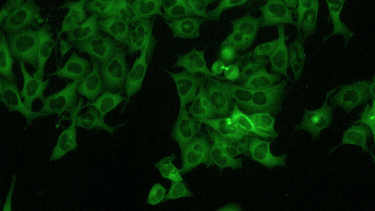
DMIL Fluorescence for Fluorescence Cell CultureThe DM IL LED is tailored for phase contrast and fluorescence monitoring of cell cultures and individual cells. With its advanced LED illumination system and precision optics, it supports routine tasks in cell culture labs like confluence estimation and transfection efficiency checks. Elevate your cell culture research with the DM IL LED, providing superior fluorescence capabilities for enhanced visualization and insights into the dynamic of cell cultures.
Loading...
View product details+
Product includes
Loading...
|
||||||||||||||||||||||||||||||||||||||||||||||||||||||

DMIL Fluorescence for Fluorescence Cell Culture with DocumentationThe DM IL LED, equipped with a K3C camera, is tailored for phase contrast and fluorescence imaging of cell cultures and individual cells. The system and the intuitive LASX software document seamlessly routine tasks in cell culture labs like confluence estimation and transfection efficiency checks. Elevate your cell culture research with the DM IL LED, featuring a color camera for seamless image capture and providing superior fluorescence capabilities for enhanced visualization and insights into dynamic cell culture phenomena.
Loading...
View product details+
Product includes
Loading...
|
||||||||||||||||||||||||||||||||||||||||||||||||||||||


Don’t see your Configuration? Request for individual Quote
