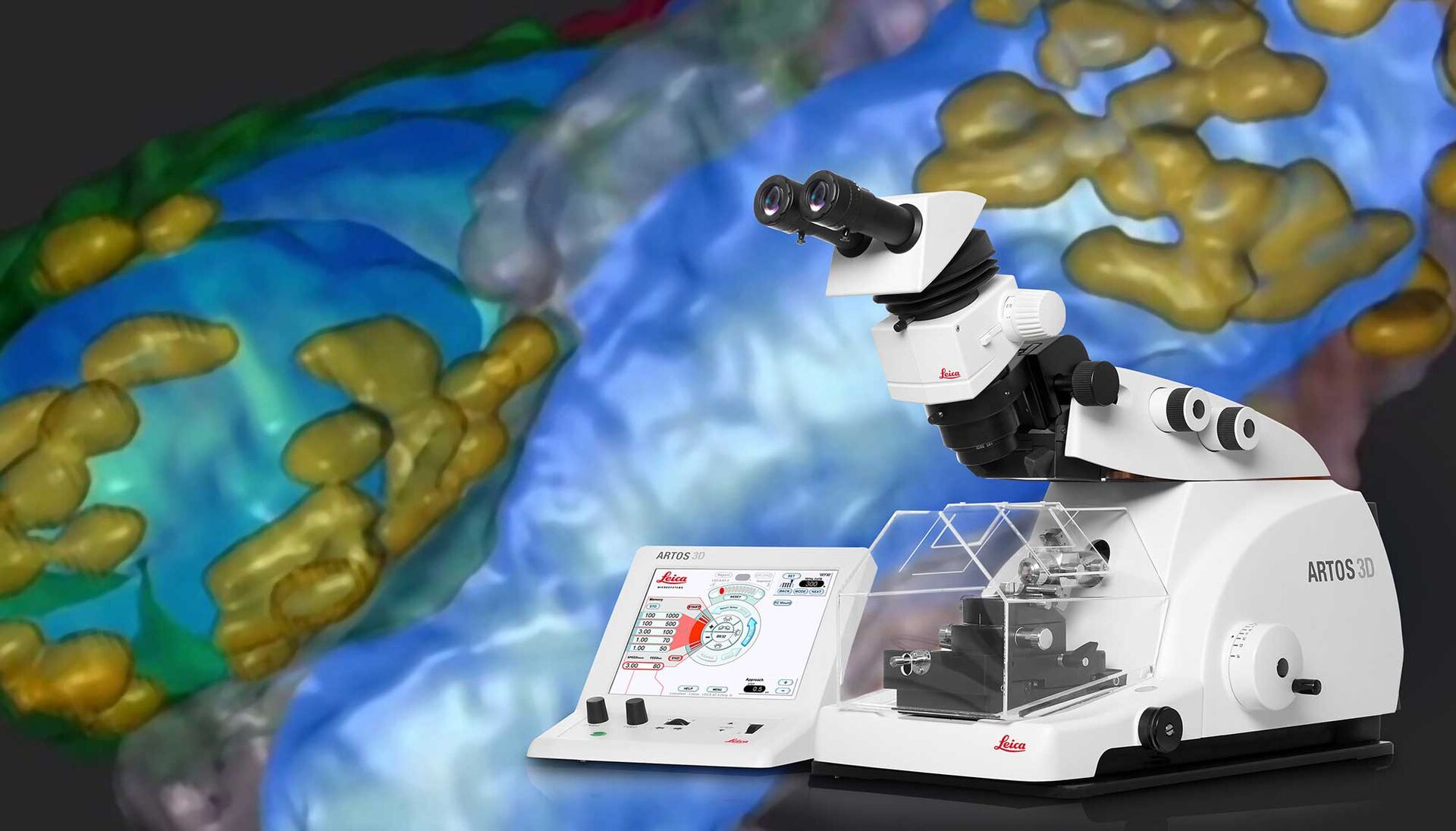
ARTOS 3D Array Tomography Solution
Get Quality Serial Sections for Array Tomography Fast
Get consistent, ultrathin serial sections for array tomography in less time with the ARTOS 3D ultramicrotome.
The ARTOS 3D (ARray TOmography Solution) automatically creates and collects hundreds of serial-section ribbons all ready for array tomography with your scanning electron microscope (SEM). Save time and effort in biological sample preparation and SEM setup procedure so you can quickly get the images you need to answer critical research questions.
- Pre-program the ARTOS 3D to produce automatically hundreds of ultrathin section ribbons with flexible block-face sizes (µm to mm)
- Avoid tricky, time-consuming manual ribbon collection with the ARTOS 3D integrated collection of fully aligned ribbons
- Save SEM setup time by loading several carriers with high section density simultaneously
- Smoothly transfer the section carrier through the whole specimen preparation process for a streamlined workflow
As transparent section carriers are available, the ARTOS 3D is also an ideal solution for correlative light and electron microscopy (CLEM).
For research use only.
Faster Array Tomography
How does the ARTOS 3D ultramicrotome allow you to save time when making high quality sections for array tomography?
Array tomography (AT) is a 3D image reconstruction technique for high resolution, quantitative analysis of biological structures. For optimal results, ultrathin, ordered sections are a pre-requisite.
Until now, AT has involved several time-consuming and cumbersome manual steps. The ARTOS 3D solution speeds up the process by automating specimen sectioning and minimizing the time required to align the sections for SEM imaging.
Read on to find out how the ARTOS 3D helps you speed up the workflow while also achieving high quality results.

Fast Sectioning & Easy Alignment for Imaging ARTOS 3D vs. Conventional Ultramicrotome
Less Work for You with the ARTOS 3D Workflow
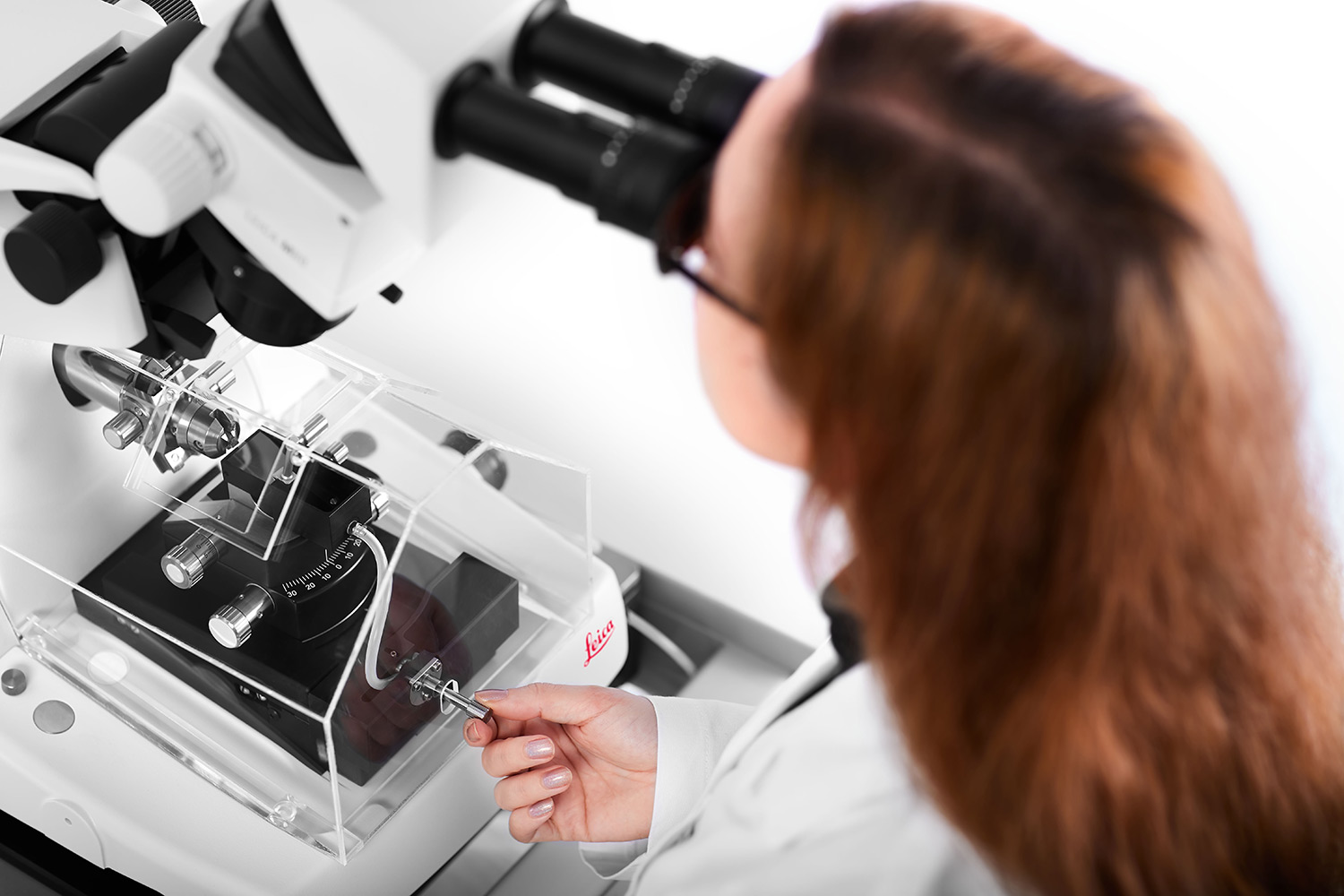
To obtain excellent results with array tomography in a timely manner, it is crucial to create ultrathin, consistent, and correctly ordered ribbons efficiently. To achieve this advantage, the ARTOS 3D ultramicrotome automates the process of making high quality, ultrathin (>20 nm) sections. The automation eliminates several of the manual work steps required by more conventional ultramicrotomes, such as the EM UC7 also from Leica Microsystems. This automation leads to a large time-savings for serial sectioning and ribbon alignment before SEM imaging.
The manual work steps required with conventional ultramicrotomes are minimized or eliminated by the ARTOS 3D system. There is:
- No manually aligning nor positioning of the ribbon sections with an eyelash
- No manual handling of the ribbons multiple times for repeat sectioning which increases greatly the risk of wrinkling
- No need to perform over and over the same tedious manual process to get wrinkle-free ribbons on the section carrier

Save Time Making High Quality Sections with a Seamless Workflow
Fast section preparation with much less effort
With the ARTOS 3D ultramicrotome, you have a seamless workflow through the whole preparation process by eliminating manual work steps:
- Fast setup with programs pre-defined by the user for different section carriers
- Automated creation and collection of hundreds of serial sections with minimal user intervention
- No more time-consuming and fiddly manual collection involving sorting and positioning of ribbons to attain proper alignment for SEM imaging

Reproducible, Artifact-Free Sections
Consistently high quality sections
The ARTOS 3D ultramicrotome is able to deliver reproducible, high quality sections rapidly by:
- Avoiding the artifact-causing conditions of manual sectioning and manipulation due to integrated direct section-ribbon collection
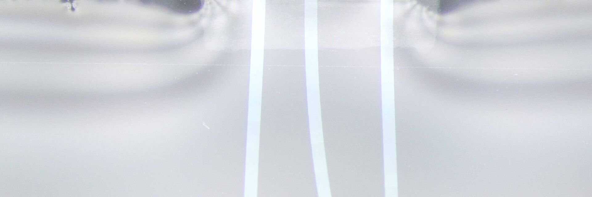
- Collecting ultrathin section ribbons without wrinkling by easily adjusting the water flow with the front valve
- Minimizing variation in section thickness by eliminating air turbulence and vibration thanks to a specially-designed draft shield and active damping plate
- Sectioning ribbons precisely with the custom-designed 4 mm diamond knife, adaptable to different section carriers
Application: 3D Image Reconstruction of Lymph Nodes
Precise section preparation with the ARTOS 3D enables optimal results with array tomography
In this application, mouse popliteal lymph nodes have been reconstructed with array tomography using serial sections produced by the ARTOS 3D solution. Array tomography enables precise examination of the cellular and protein structures of the lymph nodes, which is critical for various types of immune and cancer studies.
Learn more about how the ARTOS 3D helped optimize this array tomography application in the following report.
In addition, the consistent, reproducible sections of the ARTOS 3D ultramicrotome are also ideal for CLEM applications.
