Examining Critical Developmental Events in High-Definition
Learn how to use AI to extend live cell imaging in embryo development

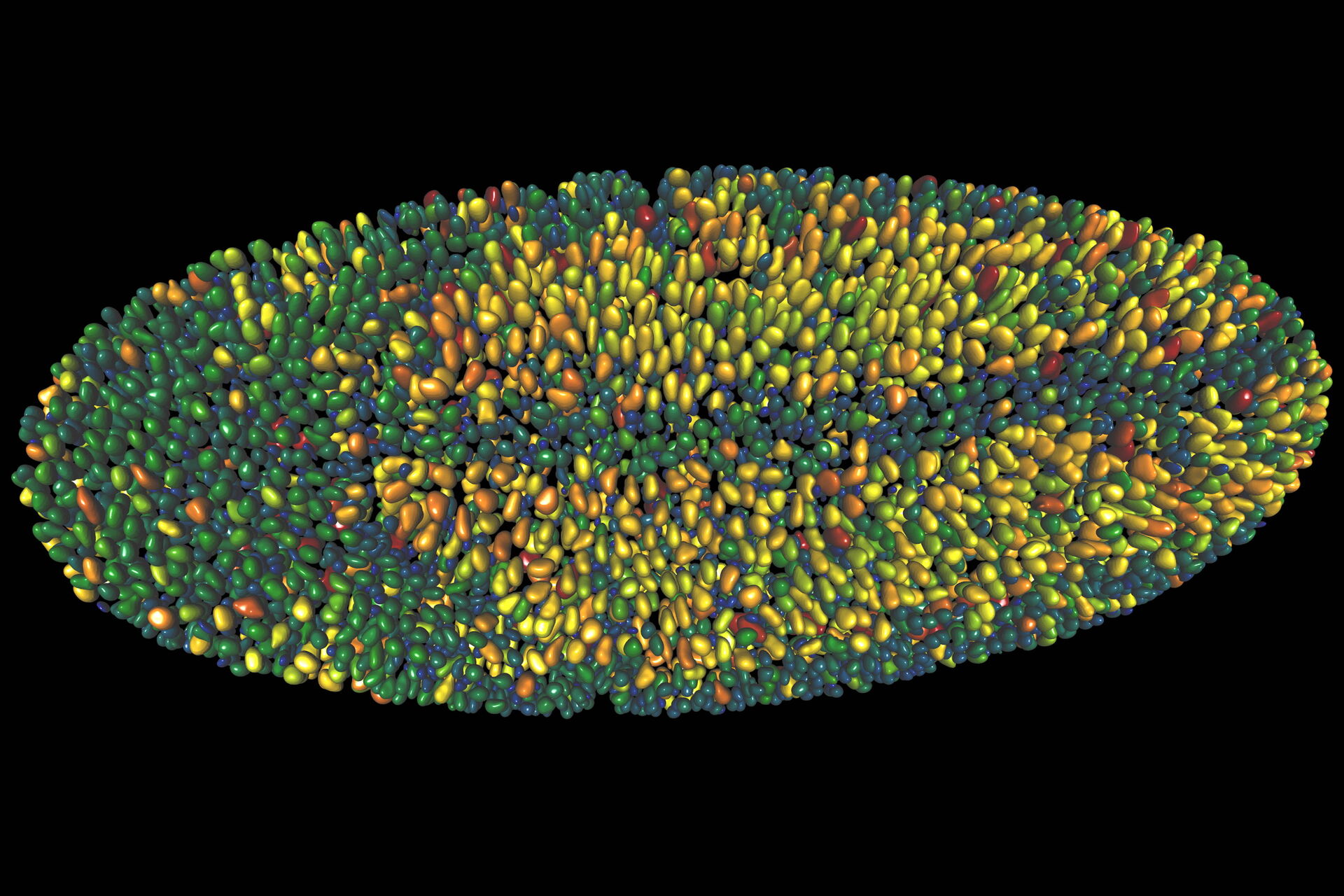
Extended live cell imaging of embryo development requires a delicate balance between light exposure, temporal resolution and spatial resolution to maintain cells’ viability. Compromises between the three factors are needed to achieve the optimal analysis outcome and to unlock greater insights from your imaging data. In this workshop, the Aivia team will demonstrate how AI can help you extend live cell imaging in embryo development.
What to expect in the webinar
Key Learnings
Researchers dealing with sample integrity challenges in developmental research can benefit from automated workflows that streamline image analysis.
- Examine critical developmental events in high-definition with minimal sample damage
- Sidestep light exposure and resolution limits to prolong time lapse imaging
- Deploy smart segmentation to easily detect objects
Related Articles
-

Overcoming Challenges with Microscopy when Imaging Moving Zebrafish Larvae
Zebrafish is a valuable model organism with many beneficial traits. However, imaging a full organism…
Jan 22, 2025Read article -

Aneurysm Clipping: Assessing Perforators in Real-Time with AR Fluorescence
This article covers two aneurysm clipping cases highlighting the clinical benefits of GLOW800…
Jan 14, 2025Read article -

Advancing Uterine Regenerative Therapies with Endometrial Organoids
Prof. Kang's group investigates important factors that determine the uterine microenvironment in…
Sep 03, 2024Read article
