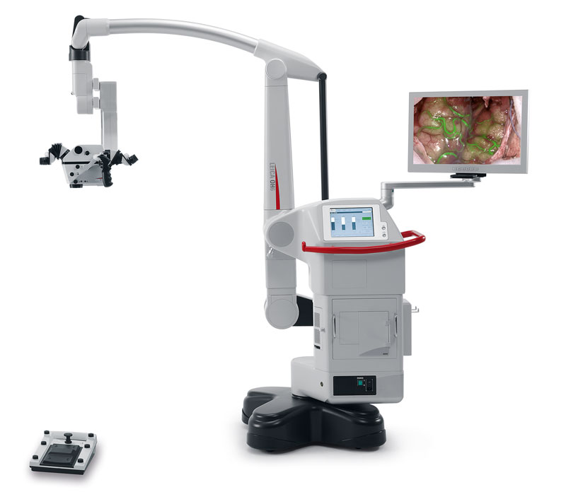
About GLOW800
With GLOW800 AR fluorescence and ICG, surgeons can observe cerebral anatomy in natural color, augmented by real-time vascular flow, with full depth perception.
Whether clipping an aneurysm, removing an AVM, performing a microvascular decompression or a bypass, surgeons can see GLOW800 directly in the eyepieces and view blood flow without interrupting workflow.
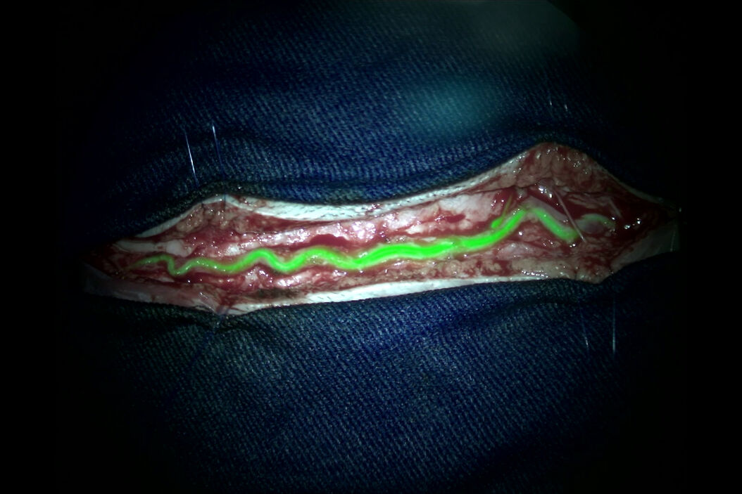
Scroll through the gallery for some impressions on GLOW800!
Dura Vessels
GLOW800 Augmented Reality Fluorescence showing dura vessels. Video courtesy of Cleopatra Charalampaki, MD, PhD, Professor of Neurosurgery, Department of Neurosurgery, Cologne Medical Center, Germany.
AVMs (Arteriovenous malformations)
Visualizing flow dynamics in an intracranial AVM, pre excision. Video courtesy of Professor Philippe Bijlenga, HUG, Geneva, Switzerland.
Visualizing flow dynamics in an ArterioVenous Fistula, pre- excision. Video courtesy of Professor Feres Chaddad, UNIFESP, Sao Paolo, Brazil.
Visualizing flow dynamics in an arterioaenous fistula, post-excision. Video courtesy of Professor Feres Chaddad, UNIFESP, Sao Paolo, Brazil.
Visualizing flow dynamics in an arteriovenous malformation, pre-excision. Video courtesy of Prof. Raphael Guzman, Basel University Hospital, Basel, Switzerland.
Visualizing flow dynamics in an arteriovenous malformation, post-excision. Video courtesy of Prof. Raphael Guzman, Basel University Hospital, Basel, Switzerland.
Visualizing flow dynamics in an arteriovenous malformation, pre-excision. Video courtesy of Dr. T. Y. Lau, Lakeland Health Center, Florida, USA.
Visualizing flow dynamics in an arteriovenous malformation, post-excision. Video courtesy of Dr. T. Y. Lau, Lakeland Health Center, Florida, USA.
GLOW800 Augmented Reality Fluorescence showing an arteriovenous (AV) fistula. Video courtesy of Cleopatra Charalampaki, MD, PhD, Professor of Neurosurgery, Department of Neurosurgery, Cologne Medical Center, Germany.
Aneurysms
Visualizing flow dynamics in an intracranial aneurysm, pre-clipping. Video courtesy of Professor Feres Chaddad, UNIFESP, Sao Paolo, Brazil.
Visualizing flow dynamics in an intracranial aneurysm, post-clipping. Video courtesy of Professor Feres Chaddad, UNIFESP, Sao Paolo, Brazil.
Visualizing flow dynamics in an intracranial aneurysm, pre- and post-excision. Video courtesy of Prof. Raphael Guzman, Basel University Hospital, Basel, Switzerland.
Visualizing flow dynamics in an intracranial aneurysm, pre-clipping. Video courtesy of Dr Chris Nickele, Semmes Murphy Hospital, Memphis, Tennessee, USA.
Visualizing flow dynamics in an intracranial aneurysm, post-clipping. Video courtesy of Dr Chris Nickele, Semmes Murphy Hospital, Memphis, Tennessee, USA.
Visualizing flow dynamics in an intracranial aneurysm, pre- and post-excision. Video courtesy of Dr. Jacques Morcos, University of Miami Health System, Miami, Florida, USA.
GLOW800 Augmented Reality Fluorescence showing an internal carotid artery (ICA) aneurysm. Video courtesy of Cleopatra Charalampaki, MD, PhD, Professor of Neurosurgery, Department of Neurosurgery, Cologne Medical Center, Germany.
GLOW800 Augmented Reality Fluorescence showing an ICA aneurysm. Video courtesy of Cleopatra Charalampaki, MD, PhD, Professor of Neurosurgery, Department of Neurosurgery, Cologne Medical Center, Germany.
GLOW800 Augmented Reality Fluorescence showing an ICA aneurysm (pre-clipping). Video courtesy of Cleopatra Charalampaki, MD, PhD, Professor of Neurosurgery, Department of Neurosurgery, Cologne Medical Center, Germany.
GLOW800 Augmented Reality Fluorescence showing an ICA aneurysm (post-clipping). Video courtesy of Cleopatra Charalampaki, MD, PhD, Professor of Neurosurgery, Department of Neurosurgery, Cologne Medical Center, Germany.
GLOW800 Augmented Reality Fluorescence showing MCA aneurysm.
GLOW800 Augmented Reality Fluorescence in Aneurysm treatment. Video courtesy of Prof. Dr. med. Nils Ole Schmidt, Neurosurgeon, University Hospital Hamburg Eppendorf
GLOW800 Augmented Reality Fluorescence in Aneurysm treatment. Showing post-clipping assessments. Video courtesy of Prof. Dr. med. Nils Ole Schmidt, Neurosurgeon, University Hospital Hamburg Eppendorf
Bypass
Visualizing flow dynamics in an intracranial bypass procedure, showing the donor vessel. Video courtesy of Professor Feres Chaddad, UNIFESP, Sao Paolo, Brazil.
Visualizing flow dynamics in an intracranial bypass procedure, showing pre-graft anastomosis. Video courtesy of Professor Feres Chaddad, UNIFESP, Sao Paolo, Brazil.
Visualizing flow dynamics in an intracranial bypass procedure, showing post-graft anastomosis. Video courtesy of Professor Feres Chaddad, UNIFESP, Sao Paolo, Brazil.
Visualizing flow dynamics in an intracranial bypass procedure, showing pre-graft anastomosis. Video courtesy of Prof. Raphael Guzman, Basel University Hospital, Basel, Switzerland.
Visualizing flow dynamics in an intracranial bypass procedure, showing post-graft anastomosis. Video courtesy of Prof. Raphael Guzman, Basel University Hospital, Basel, Switzerland.
Visualizing flow dynamics in an intracranial bypass procedure, showing pre-bypass graft. Video courtesy of Dr. T. Y. Lau, Lakeland Health Center, Florida, USA.
Visualizing flow dynamics in an intracranial bypass procedure, showing post- bypass graft. Video courtesy of Dr. T. Y. Lau, Lakeland Health Center, Florida, USA.
GLOW800 Augmented Reality Fluorescence in Extra-Cranial to Intra-Cranial Bypass surgery. Assessing extra-cranial vessel. Video courtesy of Prof. Dr. Feres Chaddad, Head of Vascular Neurosurgery at the Federal University of Sao Paulo (UNIFESP), Sao Paulo, Brazil