
Doubling the number of distinguishable colours
Using cultured cells, common staining protocols, and commercially available fluorochromes and microscopes, we exploit that the number of fluorochromes in STED or confocal microscopy can be increased by phasor-based fluorescence lifetime imaging microscopy (FLIM) separation of two dyes with similar emission spectra but different fluorescent lifetimes.
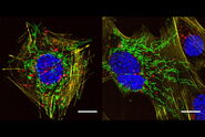
In our multicolor FLIM-STED approach, two fluorochromes in the near red (exc. 594 nm, em. 600–630 nm) and two in the far-red channel (exc. 633 nm, em. 641–680 nm), supplemented by a single further redshifted fluorochrome (exc. 670 nm, em. 701–750 nm) were all depleted with a single laser at 775 nm. The analysis, based on the phasor plot, granted an easy and quick way to visualize the quality of the stainings, the lifetime of the fluorophores, and the tools for an easy unmixing. We also provide evidence that eight-color FLIM-STED with a single depletion laser would be possible if suitable fluorochromes were identified and we confirm that a fluorochrome may have different lifetimes depending on the molecules to which it is coupled.
Generally, this approach doubles the number of distinguishable colors in laser scanning microscopy. The phasor-based analysis simplifies the whole procedure, making it possible to be used by a large number of scientists after a short training.
Related Articles
-

Extended Live-cell Imaging at Nanoscale Resolution
Extended live-cell imaging with TauSTED Xtend. Combined spatial and lifetime information allow…
Mar 05, 2024Read article -
, actin network (ATTO 647N), and nuclear pore basket (CF 680R).")
The Guide to STED Sample Preparation
This guide is intended to help users optimize sample preparation for stimulated emission depletion…
Mar 05, 2024Read article -
A Versatile Palette of Fluorescent Probes
Researchers at the Max Planck Institute for Medical Research in Heidelberg have developed a general…
Jan 24, 2022Read article
Related Pages
-

Confocal Microscopes
Our confocal microscopes for top-class biomedical research provide imaging precision for subcellular…
Visit related page
