
How is the phasor plot generated?
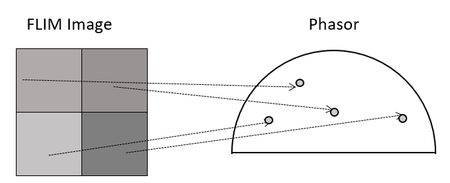
When the data is acquired with Time Resolved Single Photon Counting (TCSPC) systems, the phasor FLIM distributions are derived from a Fourier transform (Digman et al., 2008). Each pixel in the image corresponds to a point in the phasor plot.


Rules: the algebra of phasors
There are only a few rules in the phasor approach and they enable a straightforward interpretation of the lifetime distribution. Three of the most important ones are shown below (E. Gratton, 2018).
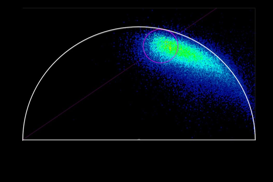
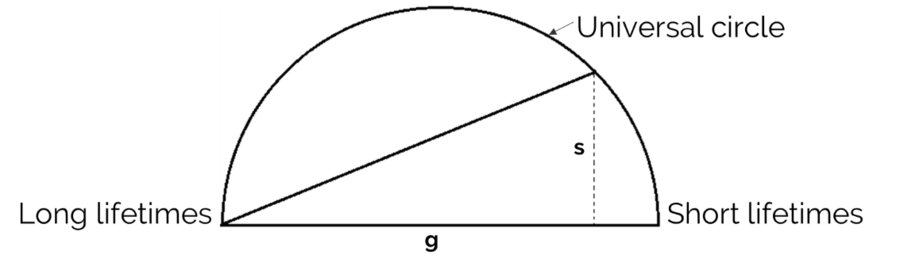
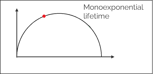
1. Single exponential lifetimes lie on the universal circle line.

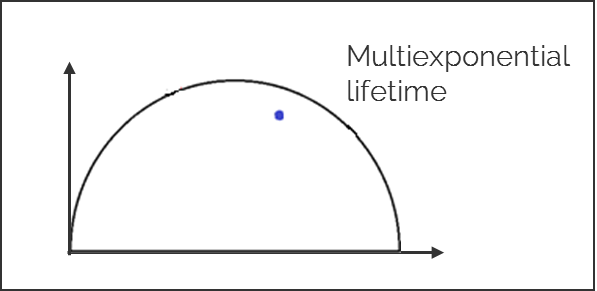
2. Multi-exponential lifetimes are found inside the universal circle and are a linear combination of their single exponential lifetime components.

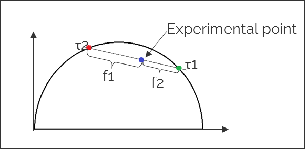
3. The ratio of the linear combination for a multi-exponential lifetime (experimentally measured) determines the fraction of the components (f1 and f2) from their two single exponential lifetimes (τ1 and τ2).

Advantages of using phasors
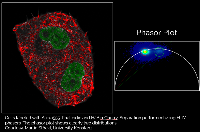
Phasor FLIM is a very powerful analysis tool for molecular species separation and FRET analysis, in particular when the donor has a multi-exponential lifetime, something which is typical of CFP [cyan fluorescent protein] (Caiolfa et al., 2007). Moreover, phasor FLIM combined with STED, allows you to use less STED power to reach the same resolution (Lanzanò et al., 2015).

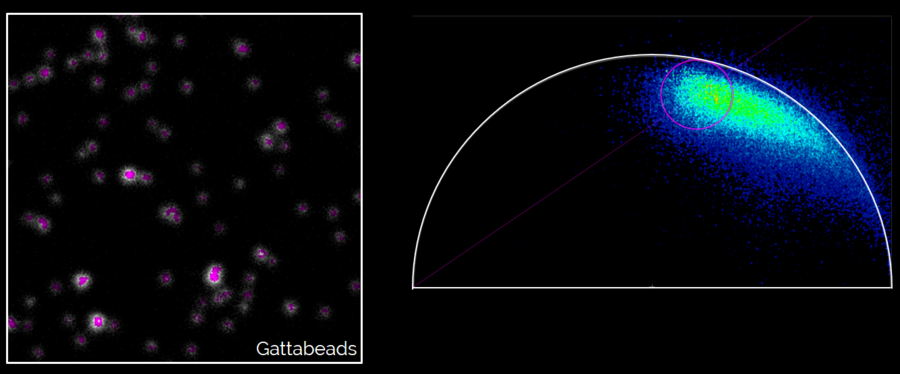
Left: Gattabeads imaged with SP8 STED system having FLIM capability. Right: By highlighting the long lifetime pixels in the phasor plot, the resolution of the STED image is improved.
References and Additional Information
- M.A. Digman, V.R. Caiolfa, M. Zamai, E. Gratton, The Phasor Approach to Fluorescence Lifetime Imaging Analysis, Biophys. J. (2008) vol. 94, iss. 2, pp. L14–L16
- V.R. Caiolfa, M. Zamai, G. Malengo, A. Andolfo, C.D. Madsen, J. Sutin, M.A. Digman, E. Gratton, F. Blasi, N. Sidenius, Monomer–dimer dynamics and distribution of GPI-anchored uPAR are determined by cell surface protein assemblies, J. Cell Biol. (2007) vol. 179, iss. 5, pp. 1067-1082
- E. Hinde, K. Yokomori, K. Gaus, K.M. Hahn, E. Gratton, Fluctuation-based imaging of nuclear Rac1 activation by protein oligomerization, Sci. Rep. (2014), vol. 4, pp. 4219
- E. Hinde, M.A. Digman, C. Welch, K.M. Hahn, E. Gratton, Biosensor FRET detection by the phasor approach to fluorescence lifetime imaging microscopy (FLIM), Microsc. Res. Tech. (2012) vol. 75, iss. 3, pp. 271–281
- L. Albertazzi, D. Arosio, L. Marchetti, F. Ricci, F. Beltram, Quantitative FRET Analysis with the E0GFP‐mCherry Fluorescent Protein Pair, Photochem. Photobiol. (2009) vol. 85, iss. 1, pp. 287-97
- L. Lanzanò, I. Hernández, M. Castello, E. Gratton, A. Diaspro, G. Vicidomini, Encoding and decoding spatio-temporal information for super-resolution microscopy, Nat. Commun. (2015) vol. 6, p. 6701
- L. Wang, B. Chen, W. Yan, Z. Yang, X. Peng, D. Lin, X. Weng, T. Ye, J. Qu, Resolution improvement in STED super-resolution microscopy at low power using a phasor plot approach. Nanoscale. (2018), vol. 10 iss. 34, pp. 16252-16260
- J. Sosnik, L. Zheng, C.V. Rackauckas, M. Digman, E. Gratton, Q. Nie, T.F. Schilling, Noise modulation in retinoic acid signaling sharpens segmental boundaries of gene expression in the embryonic zebrafish hindbrain, eLife (2016) vol. 5, e14034
- E. Gratton, The Phasor approach: Application to FRET analysis and Tissue Autofluorescence, 13th LFD workshops 2018, 22- 26 October 2018, Laboratory for Fluorescence Dynamics (LFD), University of California, Irvine, USA
- E. Gratton, The Phasor approach: Application to FRET analysis and Tissue Autofluorescence, 6th European Workshop on Advanced Fluorescence Imaging and Dynamics, 10-14 December 2018, FAB LAB, Ludwig-Maximilians-Universität München, Munich, Germany
Related Articles
-
Potential of Multiplex Confocal Imaging for Cancer Research and Immunology
Explore the new frontiers of multi-color fluorescent imaging: from image acquisition to analysis
Oct 23, 2023Read article -
Windows on Neurovascular Pathologies
Discover how innate immunity can sustain deleterious effects following neurovascular pathologies and…
Jul 04, 2023Read article -
The Power of Reproducibility, Collaboration and New Imaging Technologies
In this webinar you willl learn what impacts reproducibility in microscopy, what resources and…
Jun 27, 2023Read article
Related Pages
-
A Guide to Fluorescence Lifetime Imaging Microscopy (FLIM)
The fluorescence lifetime is a measure of how long a fluorophore remains on average in its excited…
Visit related page