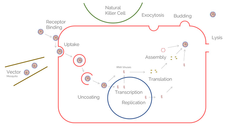
Host side of the virus infection process
For virology research model organisms are sometimes used, however, they do not mimic well the human host morphology. Hence, researchers more often rely on human tissue, biopsies, or cell culture studies.
Ex vivo infection models can be generated from tissue, biopsies, or animal models. These models can be investigated with the aid of Leica stereo microscopes.
Cell cultures, spheroids, and organoids can be monitored with the help of Leica widefield (compound) microscopes.
Further initial investigations of infection models, 2D or 3D cell cultures, are achieved with more sophisticated Leica fluorescence microscopes, such as THUNDER imagers.
More reliable downstream genetic analysis can be done with laser microdissection from Leica Microsystems which isolates specific cells from surrounding tissue.
Related articles
Microscopy in Virology

How to Sanitize a Microscope

High resolution fluorescence imaging for virology research
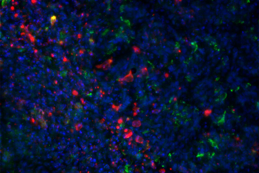
High resolution microscopy is an asset for infectious disease studies as it allows you to track causative viruses or pathogens. Combined with fluorescent labelling of cellular or viral components, it enables you to probe multiple aspects of infection processes.
Thus, both laser scanning confocal and total internal reflection fluorescence (TIRF) microscopy obtaining super-resolution are instruments of choice for viral research.
Advanced microscopy techniques, such as multiphoton and light sheet microscopy, can give you access to infection events occurring under different contexts, such as tissue sections, animal models, and 3D infection models like organoids.
Leica microscopes offering you advantages for the study of 3D specimens concerning virology research include the STELLARIS confocal platform and TIRF solutions.
Image and diagram adapted from Coomer et al. PLOS Pathogen (2020) 16(2): e1008359, DOI: 10.1371/journal.ppat.1008359

Related articles
Introduction to Fluorescent Proteins
")
How to Prepare your Specimen for Immunofluorescence Microscopy

Studying Virus Replication with Fluorescence Microscopy

How Can Immunofluorescence Aid Virology Research?
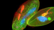
 - THUNDER Imager 3D Cell Culture Influenca virus – red, cilia – green, Nuclei – blue.")
Immersion Freezing for Cryo-Transmission Electron Microscopy: Applications

Ultrastructure of viruses
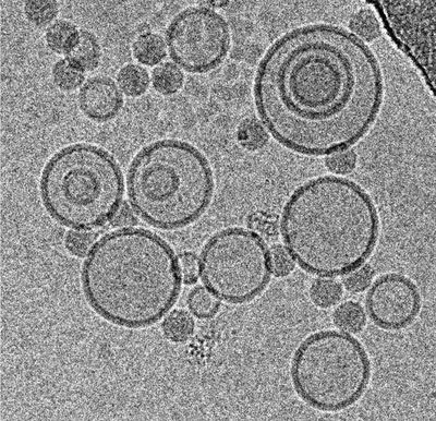
For the evaluation of virus ultrastructure, you need a resolution approaching 1 nm. At present, this resolution can only be achieved with electron microscopy. Cryo EM can achieve even sub-nanometer resolution.
Ultrastructural resolution allows you to actually see the host and virus interaction at the nanoscale, as well as confirm the results from different analytical technologies and identify potential drug target sites. Furthermore, ligands or small molecules of cell surface receptors targeted during viral particle interactions are key to the structure-guided design of vaccines and viral entry inhibitors.
For conventional and cryo EM, the sample preparation is crucial. It requires special equipment and expertise which Leica Microsystems offers you via an extensive portfolio for EM sample preparation as well as the advice from Leica experts.

 - THUNDER Imager 3D Cell Culture Influenca virus – red, cilia – green, Nuclei – blue.")