Contact Us

Museums and Art Conservation Solutions

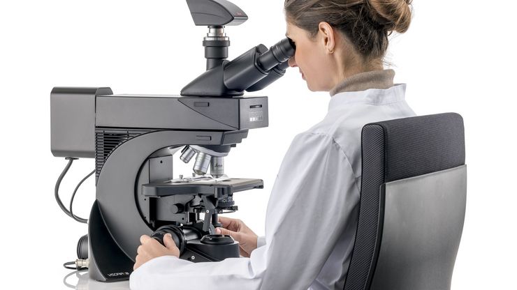
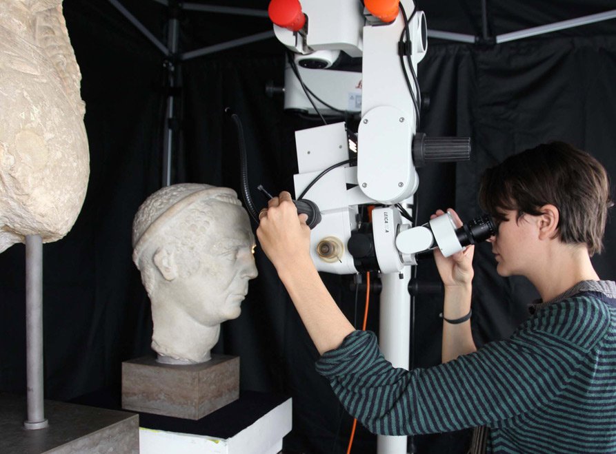
Analyzing, restoring, conserving and documenting artistic works or cultural artifacts require complex technical skills and the use of state-of-the-art imaging solutions, particularly contact-free, nondestructive methods. Combined with digital camera technology and image analysis software, Leica Microsystems’ art conservation microscope solutions are designed for the different fields of activity of restorers, art historians, archaeologists and experts in conservation workshops and museums. All microscopy solutions optimize detail-accurate work on the specimen, microscopic analysis of structures and materials as well as documentation.

Contact Us For Personal Support

Please contact us if you would like to have personal expert advice on our microscopy solutions for Art Conservation

So that you never miss a detail

Leica Microsystems provides a range of solutions for the different fields of activity of restorers, art historians, archaeologists and experts from related fields. All optimize detail-accurate work on the specimen, microscopic analysis of structures and materials as well as documentation.

Expertise for experts

Microscope solutions specially adapted to the specific requirements, combined with digital camera technology and image analysis software from Leica Microsystems, are among the professional tools used today by many experts in conservation workshops and museums.

To give you an overview of the various methods and technologies for each application area, we have listed the products that support you in your work with valuable treasures of art and irreplaceable originals.
Pigment Analysis

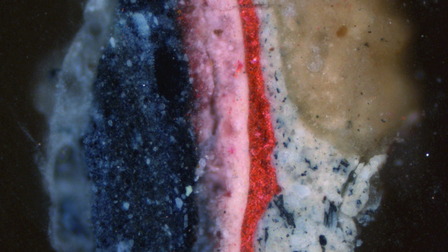
When you examine paint layers and paint pigments, you need high-resolution, high-performance microscopes with brilliant, cool illumination. Leica Microsystems provide powerful microscopy solutions with fluorescence illumination options combined with easy-to-use analysis software for optimal investigation, analysis and documentation.
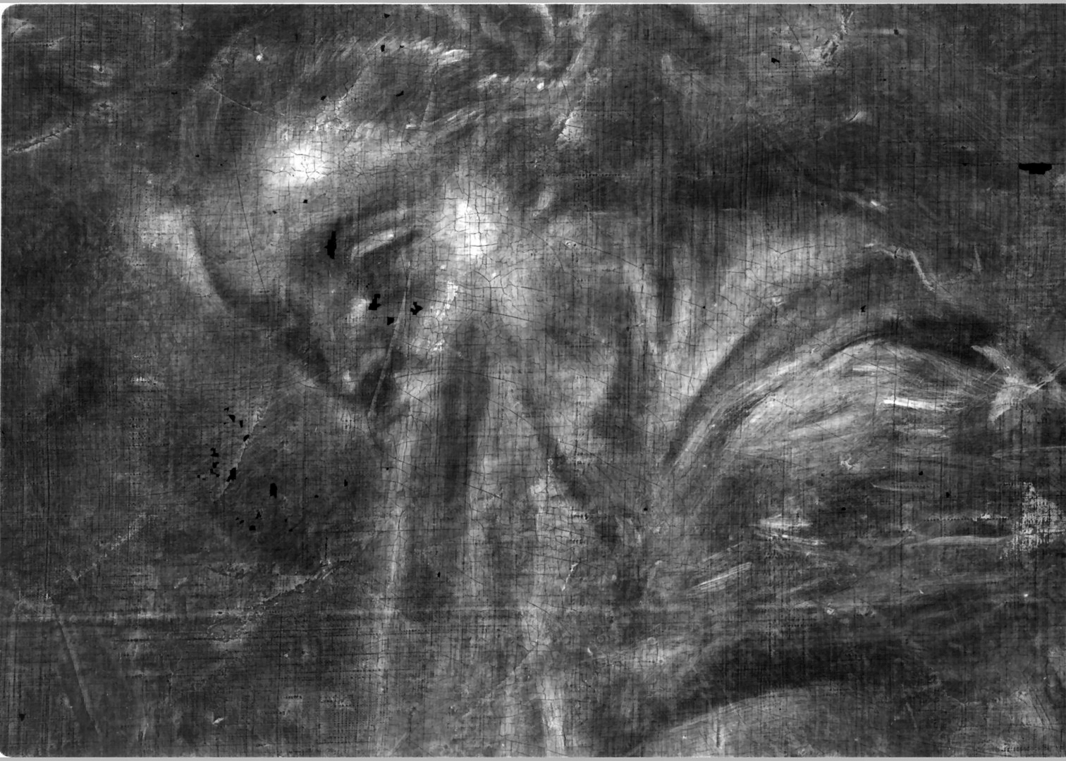
X-radiograph (detail) with the revealed head of a woman painted over.

Paintings

Detail-accurate cleaning and restoration of paintings involve the use of precise digital and stereomicroscopes customized for a variety of painting types and sizes. Leica’s microscopes for paint cleaning and restoration provide variable zoom positions which enable you to work at low magnifications and analyze the tiniest details. Floor stand options are available for larger paintings.

materials

Minerals & Fossils

Mineral and fossil conservation requires detailed image processing and analysis of through high-performance stereo and research microscopes. Whether you need to view microstructures in 3D or you need to analyze thin sections, Leica Microsystems offers versatile microscopy solutions to meet your specific application needs.

Paper, Books & Photos

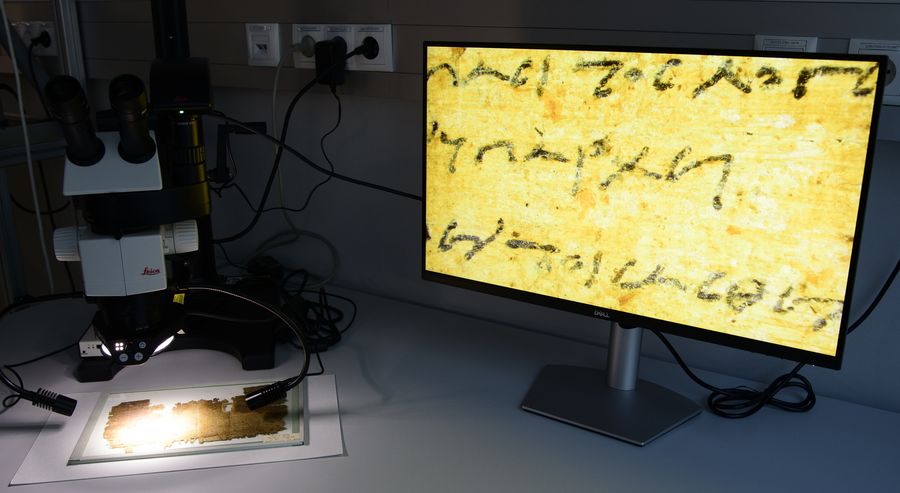
Reveal and analyze unique details of printing and finishing techniques, manuscripts or retouches and view the finest paper structures in 3D with Leica’s range of microscopy solutions. These innovative digital and stereomicroscopes combine maximum resolution and the greatest depth of field to support the acquiring, storing, annotating and documenting you need to research and restore these precious items of our heritage.

Glass, Ceramics & Stone

When you examine, clear or reconstruct glass, ceramics or stone, you need customized, high-resolution microscope solutions that enable you to analyze objects of different sizes and shapes. Leica Microsystems provides a variety of stereo and research microscopes with different illumination methods and portable stand systems tailored to your needs.

Metal

Examine signs of corrosion, dirt deposits, treatment or processing techniques for metal objects or analyzing the metal structure and determining alloy components with Leica Microsystems' individualized and affordable system solutions. Featured stereo and research microscopes are combined with digital cameras and easy-to-use 3D software to support your analysis, cleaning and documentation of metal objects.

Wood

Clean and restore painted or varnished wood objects and determine the structure and components of the applied layers with Leica's stereo and research microscope solutions. These flexible solutions are combined with table or floor stands, digital cameras and easy-to-use analysis software to support your wood conservation work.
Dutch cabinet

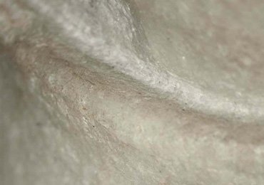
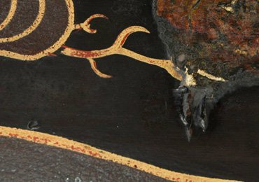
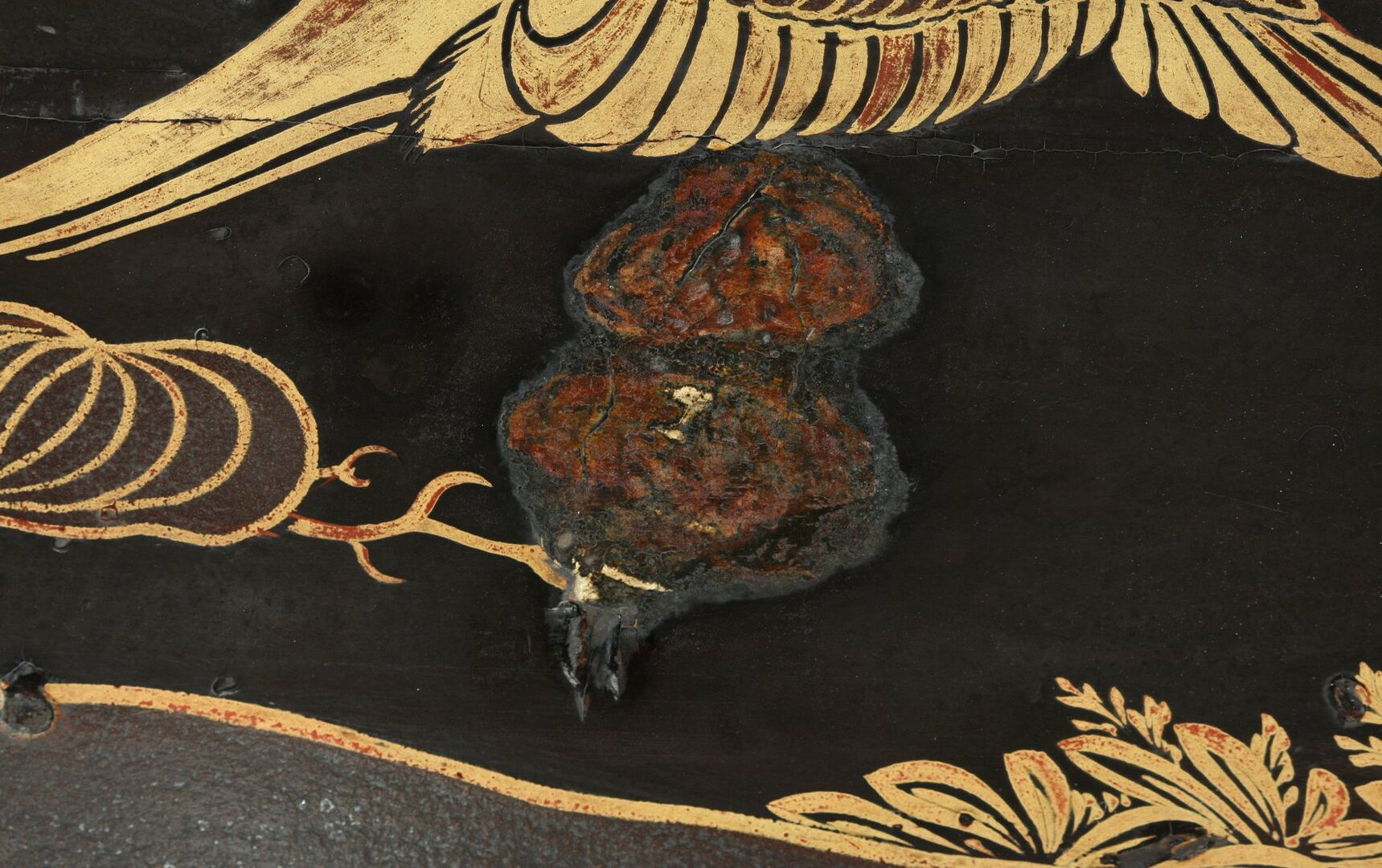
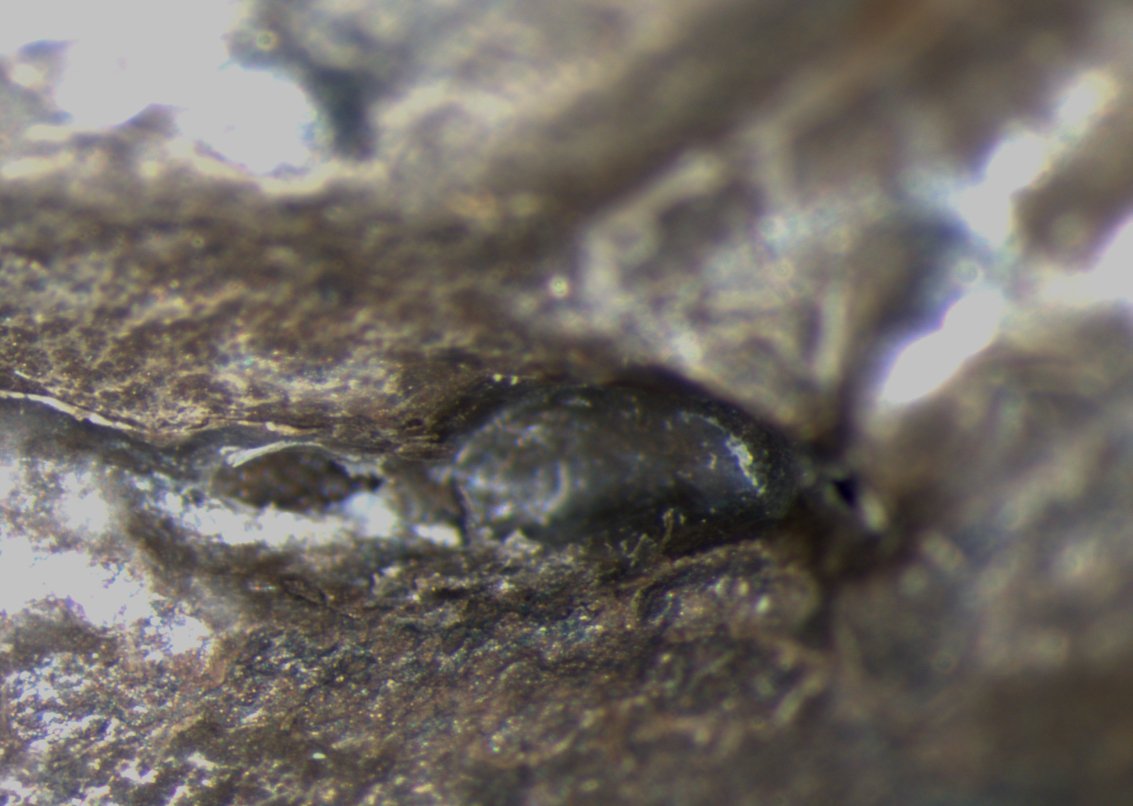
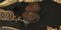
Late 17th century Dutch cabinet decorated with Japanese lacquer panels and inlaid with a motif known as seaweed marquetry. A stereomicroscope was used to analyze and clean the surface, to identify damage, and to take and analyze samples. With the help of a floor stand even areas that are difficult to access can be investigated. Damages to the cabinet were analyzed under the microscope and documented with a digital camera. This area of damage was covered with a painting of a flower. This was done in Europe, probably with oil paint. Since they form part of the history of the object, it was decided not to remove these overpaintings.

© Rijksmuseum Amsterdam

Silver candelabra

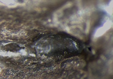
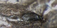
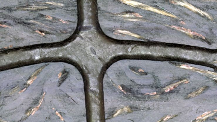
For restoration and conservation of metal objects, e.g. this silver candelabra from the 19th century. A microscope was used for analyzing damaged areas, cleaning the surface, and documentation. Under the microscope an artificial patina was discovered in the tarnished areas of the silver candelabra.

© Rijksmuseum Amsterdam

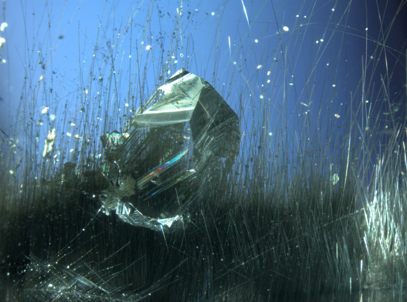
Rock crystal (quartz) and rutile needles enclosed in rock crystal (quartz), from Minas Gerais, Brazil. Because of the light metallic lustre of the rutile needles (titanium oxide, TiO2), the trade name of this quartz variety is “platinum quartz”. The picture shows a first generation rock crystal inclusion that was enclosed by a second generation of quartz. Although the inclusion and the host material have the same refractive index, the enclosed quartz is easily recognizable due to a thin film of air at the interfaces. Width of image: approx. 6 mm, transmitted light, crossed polarizers, first-order red compensator. © Michael Hügi

Rock crystal

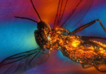
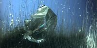
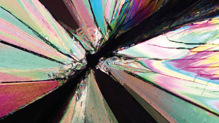
Rock crystal (quartz) and rutile needles enclosed in rock crystal (quartz), from Minas Gerais, Brazil. Because of the light metallic lustre of the rutile needles (titanium oxide, TiO2), the trade name of this quartz variety is “platinum quartz”. The picture shows a first generation rock crystal inclusion that was enclosed by a second generation of quartz. Although the inclusion and the host material have the same refractive index, the enclosed quartz is easily recognizable due to a thin film of air at the interfaces. Width of image: approx. 6 mm, transmitted light, crossed polarizers, first-order red compensator.

© Michael Hügi

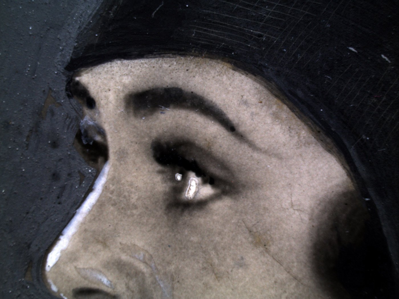
For restoration of ancient photos, high-end stereomicroscopes combined with digital cameras are used for acquiring, storing, annotating and displaying high quality images of our heritage which needs restoration. The microscopic analysis supports the recognition of the original material, the analysis of the conservation status and effects of different methods of restoration. © Alinari, Florence

Ancient photos

For restoration of ancient photos, high-end stereomicroscopes combined with digital cameras are used for acquiring, storing, annotating and displaying high quality images of our heritage which needs restoration. The microscopic analysis supports the recognition of the original material, the analysis of the conservation status and effects of different methods of restoration.

© Alinari, Florence

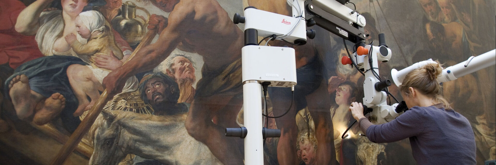
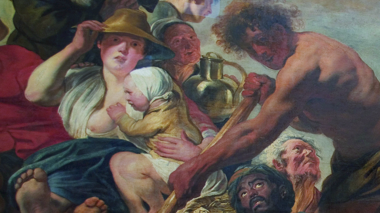
Jacob Jordaens (1593–1678), “The Tribute Money. Peter Finding the Silver Coin in the Mouth of the Fish”, also called “The Ferry Boat to Antwerp” (Oil on canvas, 279.5 by 467 cm. With a stereomicroscope combined with a floor stand the paint surface has been investigated and analyzed. © Statens Museum for Kunst, Copenhagen

“The Ferry Boat to Antwerp”

Jacob Jordaens (1593–1678), “The Tribute Money. Peter Finding the Silver Coin in the Mouth of the Fish”, also called “The Ferry Boat to Antwerp” (Oil on canvas, 279.5 by 467 cm. With a stereomicroscope combined with a floor stand the paint surface has been investigated and analyzed.

© Statens Museum for Kunst, Copenhagen

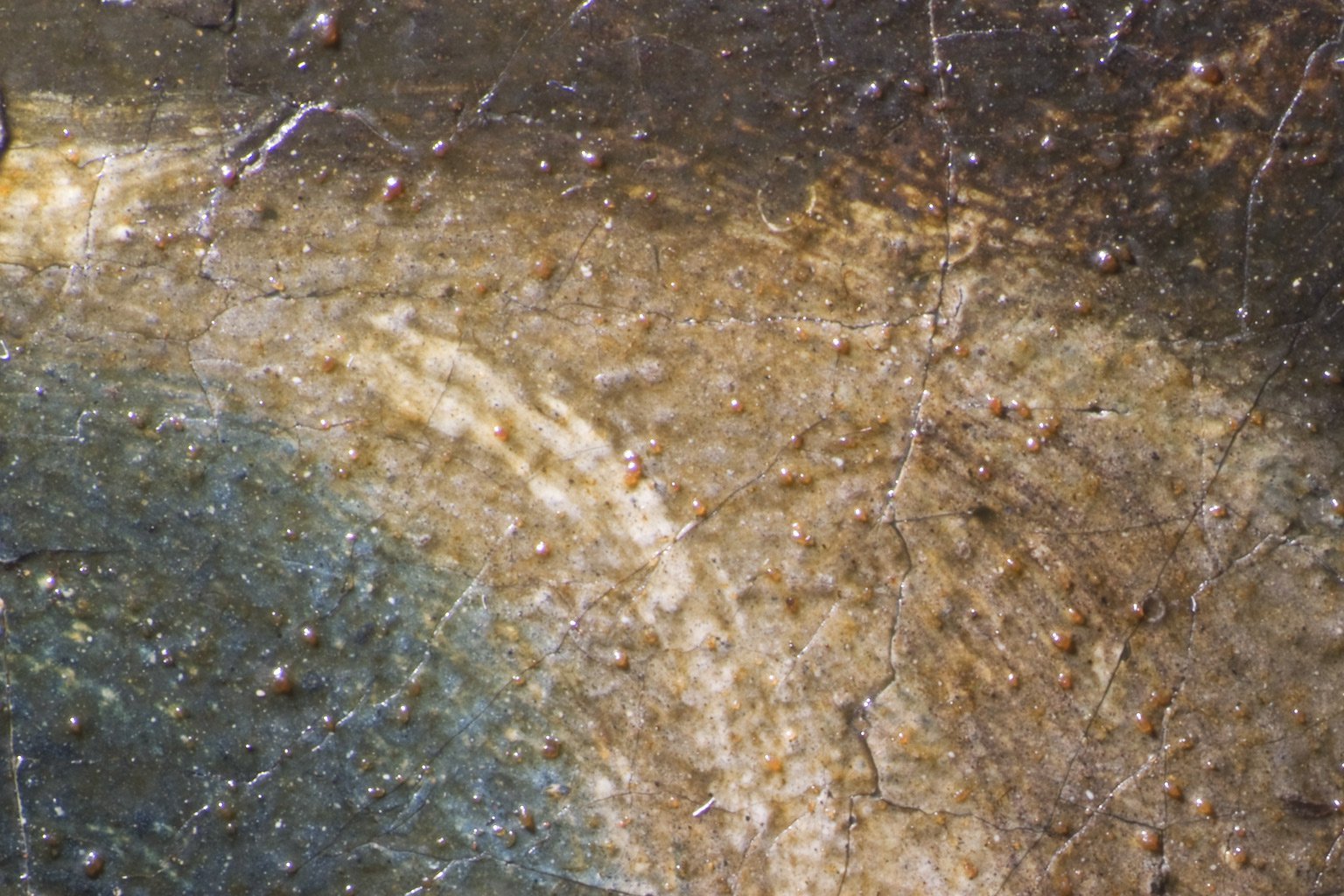
Willem van Honthorst (1594-1666) “Portrait of a Young Man Holding a Plumed Hat in his Right Hand” The almost life-size portrait dating from the first half of the 17th century requires extensive restoration. The microscope examination shows the rough granular surface of this Dutch painting which is a result of a later application of varnish with poorly dissolved components. ©Sammlungen des Fürsten von und zu Liechtenstein, Vaduz–Wien

“Portrait of a Young Man Holding a Plumed Hat in his Right Hand”

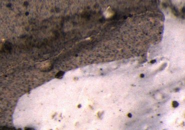
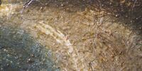
Willem van Honthorst (1594-1666) “Portrait of a Young Man Holding a Plumed Hat in his Right Hand”; The almost life-size portrait dating from the first half of the 17th century requires extensive restoration. The microscope examination shows the rough granular surface of this Dutch painting which is a result of a later application of varnish with poorly dissolved components.

© Sammlungen des Fürsten von und zu Liechtenstein, Vaduz–Wien

Rock crystal (quartz) and rutile needles enclosed in rock crystal (quartz), from Minas Gerais, Brazil. Because of the light metallic lustre of the rutile needles (titanium oxide, TiO2), the trade name of this quartz variety is “platinum quartz”. The picture shows a first generation rock crystal inclusion that was enclosed by a second generation of quartz. Although the inclusion and the host material have the same refractive index, the enclosed quartz is easily recognizable due to a thin film of air at the interfaces. Width of image: approx. 6 mm, transmitted light, crossed polarizers, first-order red compensator. © Michael Hügi
For restoration of ancient photos, high-end stereomicroscopes combined with digital cameras are used for acquiring, storing, annotating and displaying high quality images of our heritage which needs restoration. The microscopic analysis supports the recognition of the original material, the analysis of the conservation status and effects of different methods of restoration. © Alinari, Florence
Jacob Jordaens (1593–1678), “The Tribute Money. Peter Finding the Silver Coin in the Mouth of the Fish”, also called “The Ferry Boat to Antwerp” (Oil on canvas, 279.5 by 467 cm. With a stereomicroscope combined with a floor stand the paint surface has been investigated and analyzed. © Statens Museum for Kunst, Copenhagen
Willem van Honthorst (1594-1666) “Portrait of a Young Man Holding a Plumed Hat in his Right Hand” The almost life-size portrait dating from the first half of the 17th century requires extensive restoration. The microscope examination shows the rough granular surface of this Dutch painting which is a result of a later application of varnish with poorly dissolved components. ©Sammlungen des Fürsten von und zu Liechtenstein, Vaduz–Wien

Related Articles

Read our latest articles about Art Conservation

The knowledge portal of Leica Microsystems offers scientific research and teaching material on the subjects of microscopy. The content is designed to support beginners, experienced practitioners and scientists alike in their everyday work and experiments.

More Articles
Images of the same area of a processed wafer taken with standard (left) and oblique (right) brightfield illumination using a Leica compound microscope. The defect on the wafer surface is clearly more visible with oblique illumination.

Rapid Semiconductor Inspection with Microscope Contrast Methods

Semiconductor inspection for QC of materials like wafers can be challenging. Microscope solutions that offer several contrast methods enable fast and reliable defect detection and efficient workflows.
Preparation of an IC-chip cross section: grinding and polishing of the chip cross section.

Cross-section Analysis for Electronics Manufacturing

This article describes cross-section analysis for electronics concerning quality control and failure analysis of printed circuit boards (PCBs) and assemblies (PCBAs), integrated circuits (ICs), etc.
Image of a Siemens star, where the diameter of the 1st black line circle is 10 mm and the 2nd is 20 mm, taken via an eyepiece of a M205 A stereo microscope. The rectangles represent the field of view (FOV) of a Leica digital camera when installed with various C-mounts (red 0.32x, blue 0.5x, green 0.63x).

Understanding Clearly the Magnification of Microscopy

To help users better understand the magnification of microscopy and how to determine the useful range of magnification values for digital microscopes, this article provides helpful guidelines.
Stereo microscopes are often considered the workhorses of laboratories and production sites.

Key Factors to Consider When Selecting a Stereo Microscope

This article explains key factors that help users determine which stereo microscope solution can best meet their needs, depending on the application.
The Emspira 3 digital microscope offers what users need for comprehensive visual inspection, including comparison, measurement, and documentation sharing.

Digital Inspection Microscope for Industrial Applications

Factors users should consider before choosing a digital inspection microscope for industrial applications, including quality control (QC), failure analysis (FA), and R&D, are described in this…
Inspection microscope image of a printed circuit board (PCB) taken with a ring light (RL) and near vertical illumination (NVI).

Microscope Illumination for Industrial Applications

Inspection microscope users can obtain information from this article which helps them choose the optimal microscope illumination or lighting system for inspection of parts or components.
Sport Fabrics Taped I3 10x Color

How Industrial Applications Benefit from Fluorescence Microscopy

Watch this free webinar to know more about what you can do with fluorescence microscopy for industrial applications. We will cover a wide range of investigations where fluorescence contrast offers new…
Visual inspection of a PCBA with the Ivesta 3 Greenough stereo microscope.

How to Select the Right Solution for Visual Inspection

This article helps users with the decision-making process when selecting a microscope as a solution for routine visual inspection. Important factors that should be considered are described.

How to Use a Digital Microscope to Streamline Inspection Processes

Watch this webinar for inspiration and expert advice on how to make quality control simpler, quicker, and easier. Learn how to perform comprehensive visual inspection, including comparison,…

Introduction to 21 CFR Part 11 and Related Regulations

This article provides an overview of regulations and guidelines for electronic records (data entry, storage, signatures, and approvals) used in the USA (21 CFR Part 11), EU (GMP Annex 11), and China…
Background image
Scroll to top