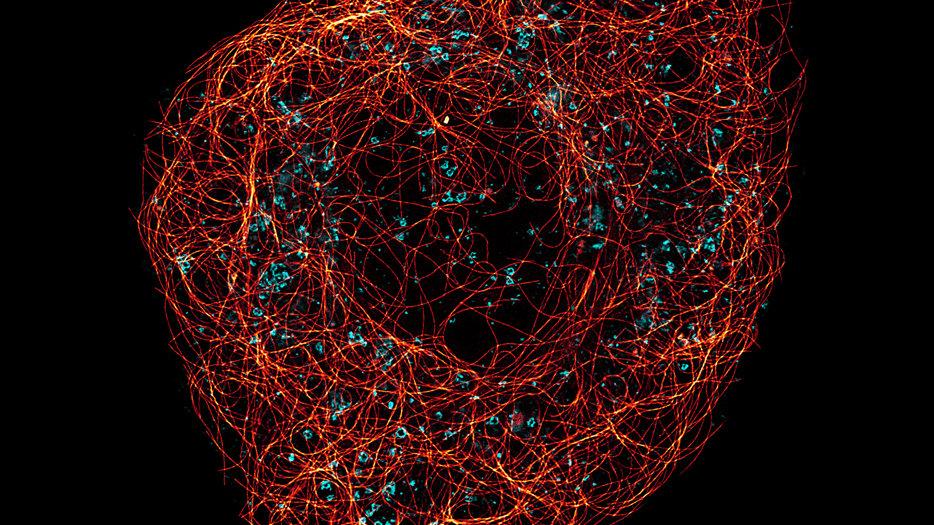
Contact Us

Super-Resolution Microscopy

Super-resolution light microscopy empowers you to study subcellular structures and dynamics with greater detail. While the spatial resolution of confocal image acquisition is doubled with the LIGHTNING technology, when using STED it can deliver insights at the nanoscale. Learn more here about Leica super-resolution methods and how they enable novel discoveries in the fields of virology, immunology, neuroscience, and cancer research.

Need Assistance?

Contact a local specialist for advice on the right super-resolution or nanoscopy system for your needs.

About Super-Resolution

Due to the diffraction limit of light, “traditional” fluorescence microscopy techniques cannot resolve structures below 240 nm. Because biology does not stop at this scale, so-called super-resolution microscopy techniques are indispensable. LIGHTNING and STED are two compelling Leica super-resolution confocal methods for visualizing structures with greater precision and uncovering details that would otherwise not be visible.

The LIGHTNING detection concept is based on adaptive deconvolution and effectively doubles the lateral resolution to 120 nm. With LIGHTNING, you can probe specimens from microtubule dynamics to the structure of subcellular compartments.

STED nanoscopy takes super resolution to the next level, enabling that you routinely achieve lateral resolutions below 30 nm. With STED, you can dissect cellular structure and function in vivo, such as the re-organization of chromatin and nuclear pores, the finest changes in neuronal structure, intracellular transport of mitochondria and synaptic vesicles, the entry of virus particles, the colocalization of protein complexes, and more.

Frequently Asked Questions Super Resolution

Show answer What is super-resolution microscopy?

Super-resolution microscopy is an optical imaging technology that overcomes the diffraction limit of light and allows the visualization of subcellular structures and dynamics in greater detail than can be achieved with conventional optical microscopy. A resolution of 30 nm is possible using STED (stimulated emission depletion) with nanoscopy. A nanoscale resolution reveals information about subcellular structures and interactions with unprecedented detail. Super-resolution microscopy enables groundbreaking discoveries in the fields of virology, immunology, neuroscience, and cancer research.

For more information about super-resolution microscopy, refer to the Science Lab articles mentioned above or go to: The Microscopy Knowledge Portal

Show answer How does super-resolution microscopy work?

Super-resolution microscopy can be performed in several ways. Here are the 3 more common methods offered by Leica Microsystems:

  1. The LIGHTNING detection concept enables super-resolution confocal microscopy. Unlike traditional deconvolution techniques that use a global set of parameters for the full image, it is based on an adaptive deconvolution process which uses a voxel-specific decision mask for finding the optimal deconvolution parameters . Thanks to powerful GPU computing, LIGHTNING works in near real-time for multiple colors simultaneously. In combination with a resonant scanner, it also provides a large field of view and high frame rate. For more information about LIGHTNING, refer to the following: Obtain maximum information from your specimen with LIGHTNING & Image information extraction by adaptive deconvolution
  2. The STED method along with confocal also allows super-resolution fluorescence microscopy. STED works with multiple channels and approaches isotropic super-resolution imaging in three dimensions. STED is based on the illumination of a diffraction-limited spot with a fluorophore-exciting wavelength and simultaneous illumination of a ring-shaped area with wavelengths that cause de-excitation or stimulated emission. This arrangement renders only a sub-diffraction area in the excited state and consequently allows probing with higher resolution than that of typical diffraction-limited optical microscopy. The unique TauSTED functionality from Leica Microsystems even allows for the increased STED resolution while eliminating undesired background noise .

For more information about STED, refer to: Video Talk on Super-Resolution: Overview and Stimulated Emission Depletion (STED) Microscopy & TauSTED: pushing STED beyond its limits with lifetime

Show answer What is the difference: super-resolution microscopy vs electron microscopy?

Whereas super-resolution microscopy exploits visible light optics to visualize a sample, electron microscopy exploits a beam of electrons for imaging a sample. Both techniques have their pros and cons, yet only super-resolution microscopy offers the advantages of experimenting with living specimens, labeling multiple molecular targets to give them specific contrast, and convenient sample preparation.

Life Science Research

Leica Microsystems’ life science research microscopes support the imaging needs of the scientific community with advanced innovation and technical expertise for the visualization, measurement and analysis of microstructures.

Read More

Virology

Do your research interests focus on viral infection and disease? Find out how you can gain insights into virology with solutions for imaging and sample preparation from Leica Microsystems.

Read More

Neuroscience

Are you working towards a better understanding of neurodegenerative diseases or studying the function of the nervous system? See how you can make breakthroughs with imaging solutions from Leica Microsystems.

Read More

Cancer Research

Cancer is a complex and heterogeneous disease caused by cells deficient in growth regulation. Genetic and epigenetic changes in one or a group of cells disrupt normal function and result in autonomous, uncontrolled cell growth and proliferation.

Read More

Related Articles

Read our latest articles about Super-Resolution Microscopy

The knowledge portal of Leica Microsystems offers scientific research and teaching material on the subjects of microscopy. The content is designed to support beginners, experienced practitioners and scientists alike in their everyday work and experiments.

More Articles
Multicolor fixed STED image. Inner ear section, mouse, TauSTED Xtend 589 on AF488 and TauSTED Xtend 775 on AF633-Phalloidin. Sample courtesy of Dennis Derstrof, Klinik für Hals-, Nasen und Ohrenheilkunde, Universität Marburg & Prof. Dr. Dominik Oliver aus dem Institut für Physiologie und Pathophysiologie, Abteilung für Neurophysiologie, Universität Marburg.

Extended Live-cell Imaging at Nanoscale Resolution

Extended live-cell imaging with TauSTED Xtend. Combined spatial and lifetime information allow super-resolution microscopy at extremely low light dose.
Multicolor TauSTED Xtend 775 for Cell Biology applications that require nanoscopy resolution for multiple cellular components. Cells showing vimentin fibrils (AF 594), actin network (ATTO 647N), and nuclear pore basket (CF 680R). Sample courtesy of Brigitte Bergner, Mariano Gonzales Pisfil, Steffen Dietzel, Core Facility Bioimaging, Biomedical Center, Ludwig-Maximilians-University, Munich, Germany.

The Guide to STED Sample Preparation

This guide is intended to help users optimize sample preparation for stimulated emission depletion (STED) nanoscopy, specifically when using the TCS SP8 STED 3X nanoscope from Leica Microsystems. It…
Five-color FLIM-STED

Five-color FLIM-STED with One Depletion Laser

Webinar on five-color STED with a single depletion laser and fluorescence lifetime phasor separation.

A Versatile Palette of Fluorescent Probes

Researchers at the Max Planck Institute for Medical Research in Heidelberg have developed a general strategy to synthesize live-cell compatible fluorogenic probes, and the result are the new MaP (Max…
STED imaging of mitochondria revealed cristae dynamics during fusion. A comparison of confocal (left) and STED (right) imaging.

Mitochondrial Dynamics Quantitatively Revealed by STED Nanoscopy

Mitochondria are vital subcellular organelles, which can generate energy to support the entire lifecycle of biological cells. However, conventional fluorescence microscopy cannot provide a high enough…

Benefits of Combining STED and Lifetime

In this interview, Professor Alberto Diaspro talks about the advantages of the White Light Laser and the TauSTED capabilities of STELLARIS 8 STED. He speaks about his experience with the confocal…

Effects of Clearing Media on Tissue Transparency and Shrinkage

This study comprehensively evaluates the effects of different clearing media on tissue transparency and shrinkage by comparing freshly dissected dipteran fly brains with their cleared equivalents.…

DNA Replication in Cancer Cells

DNA synthesis can be impeded by collisions between the DNA replication machinery and co-transcriptional R-loops leading to a major source of genomic instability in cancer cells. In this paper we…

Super-Resolution Microscopy Image Gallery

Due to the diffraction limit of light, traditional confocal microscopy cannot resolve structures below ~240 nm. Super-resolution microscopy techniques, such as STED, PALM or STORM or some…

Regulators of Actin Cytoskeletal Regulation and Cell Migration in Human NK Cells

Dr. Mace will describe new advances in our understanding of the regulation of human NK cell actin cytoskeletal remodeling in cell migration and immune synapse formation derived from confocal and…
Background image
Scroll to top