Boston Innovation Hub
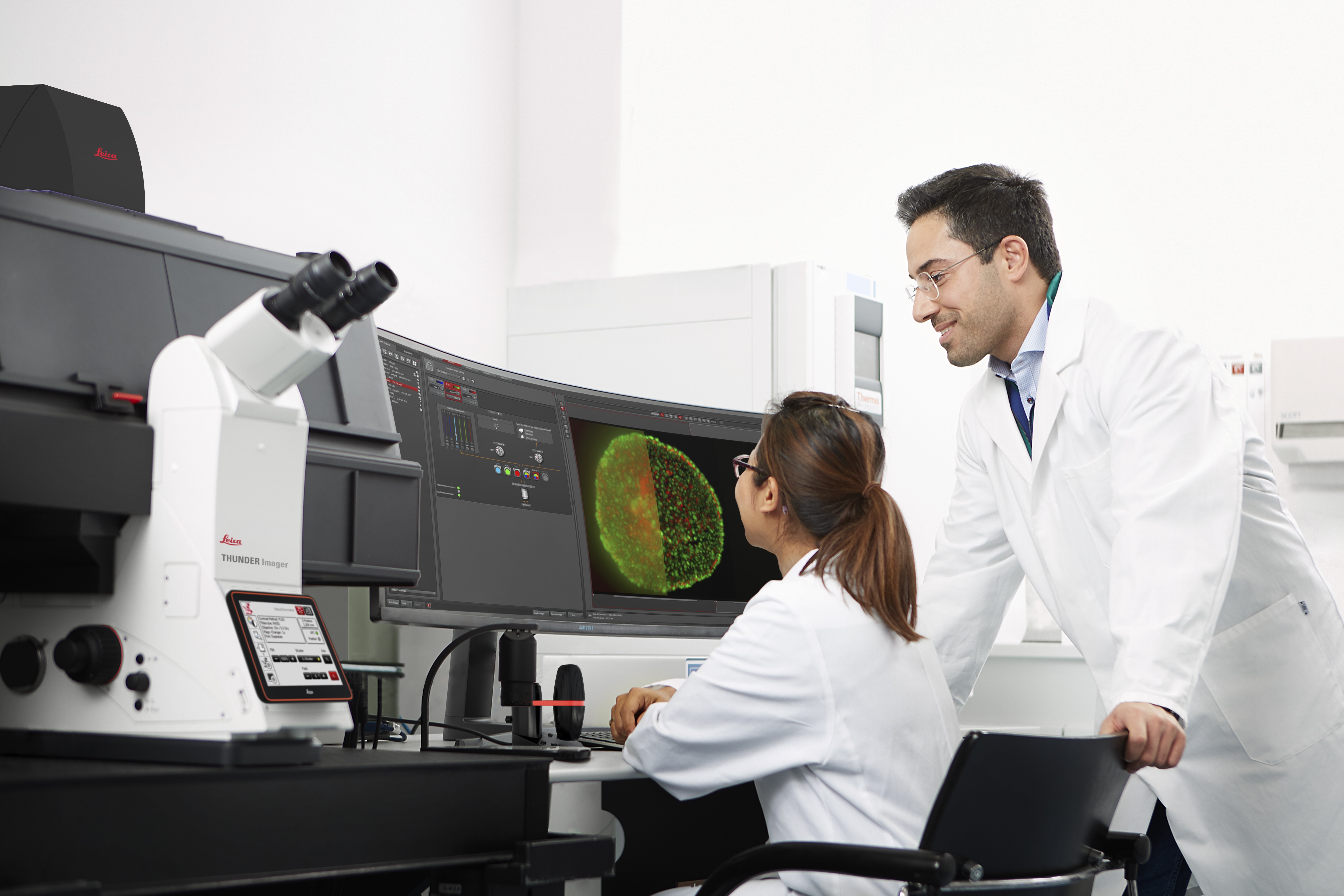
Our Boston Innovation Hub offers researchers from academia and industry access to state-of-the-art microscopy systems and support to unlock new discoveries. Our systems support the imaging needs of advanced research and provide the technical precision required to visualize dynamic events. Leica experts are on-site to empower researchers to harness data from these advanced imaging systems in order to achieve groundbreaking insights.
Experience the right imaging solutions for your applications at the Boston Innovation Hub.
MEET US AT OUR UPCOMING EVENTS
Aivia
Date: Every Tuesday
Time: 1:00 pm to 4:00 pm ET
Location: Boston Innovation Hub
Register
Level up your research through imaging
Date: Every Tuesday
Time: 1:00 pm to 4:00 pm ET
Location: Boston Innovation Hub
Register
Systems
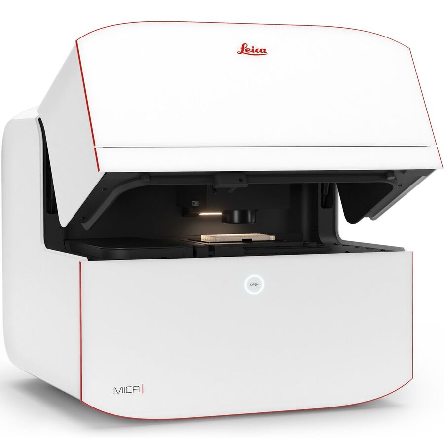
Mica
More than a highly automated microscope, Mica unites widefield and confocal imaging in a sample protecting, incubating environment. With the simple push of a button, you have everything you need - all in one place - to supercharge fluorescence imaging workflows and get meaningful scientific results faster.
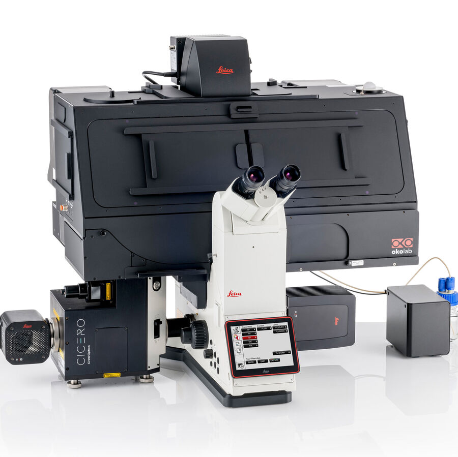
THUNDER Imager Cell Spinning Disk
Discover the power of combining THUNDER technology with the CrestOptics CICERO spinning disk.
This synergistic pairing allows you to get higher quality data and clearer details from 3D samples, delivering a deeper and more detailed view of your research.
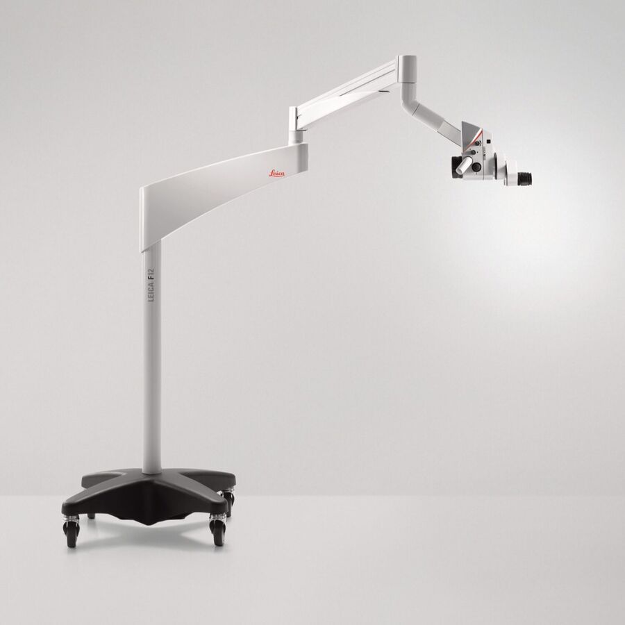
M320
- SEE WHAT YOU NEED TO SEE thanks to homogenous illumination with two integrated LED light paths providing high color perception and reproduction
- Provides the same practical, flexible, and ergonomic setup as in a medical clinic
- Record every surgery for thorough documentation using the fully integrated, cable-free 4K camera
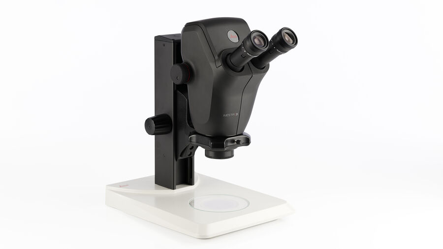
Ivesta
See the relevant details faster during inspection thanks to an optimal 3D perception which requires less adjusting of the microscope.
You can also handle the sample under the microscope objective with ease due to a large working distance.
AIVIA
Using state-of-the-art, AI-first software architecture, Aivia is a uniquely innovative and complete 2-to-5D image visualization, analysis and interpretation platform designed for the reliable processing and reconstruction of highly complex images in just minutes.
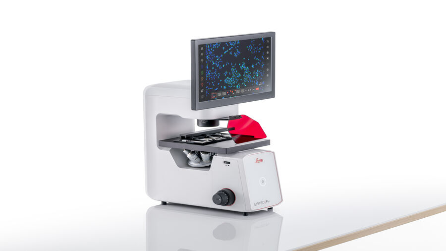
Mateo FL
Mateo FL Digital Fluorescence Microscope helps you boost your advanced cell culture research with its multi-modal fluorescence and transmitted light capabilities, automated analysis tools, and secure data tracking.
Enhance your visibility of unstained, transparent, translucent or low-contrast specimens with automated phase contrast. With a built-in dual camera system, it eliminates the need for physical camera changes and alignment, streamlining processes and improving efficiency.
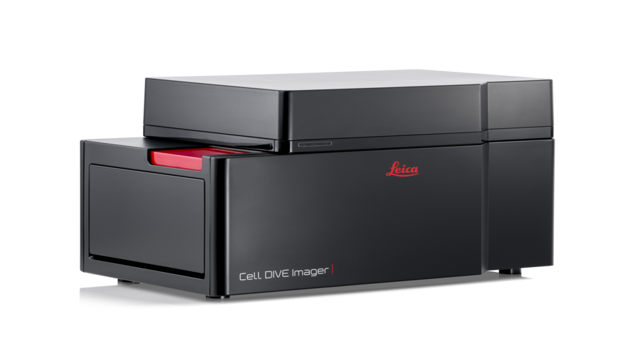
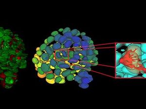
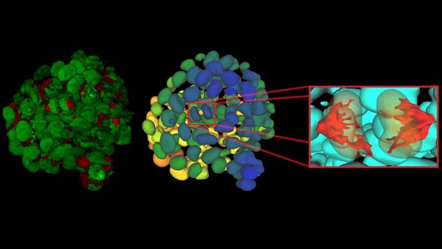
Cell DIVE Multiplex Imaging Solution
Cell DIVE is a precise, open multiplexing solution that lets your research dictate the level of automation required, which antibodies to use, how to build your antibody panel, and more.
Unveil whole tissue imaging down to the single cell level, automatically calibrated and corrected to enable quality analysis downstream.
Events
UMass Chan Medical School launches Partner in Microscopy program with Leica Microsystems
The Sanderson Center for Optical Experimentation (SCOPE) at UMass Chan Medical School and Leica Microsystems, Inc. have collaborated to establish the Partner in Microscopy site at UMass Chan. Read more...
Spatial Biology Symposium Sparks Scientific Dialogue at Boston Innovation Hub
The Boston Innovation Hub welcomed leaders from top biotech and biopharma organizations for a Spatial Biology Symposium on April 7, 2025. The symposium facilitated deep discussions on the future of spatial technologies in biological research. Read more...
TEAM
Leica Microsystems
230 3rd Ave, 4th floor, Waltham, MA 02451
lms_boston@leica-microsystems.com
+1857-331-0308
As you arrive, there is ample free parking located in the garage that is under the building, parking gates will be raised, and no tickets needed.
