


")


.")


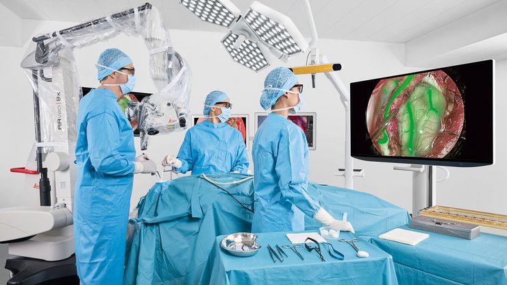
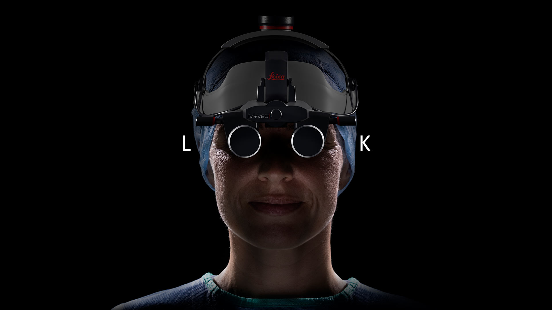
Hybrid surgical microscope empowers surgeons to perform neurosurgery, spine, and reconstructive procedures with freedom and flexibility.
Our Latest Articles
A Guide to Zebrafish Research - See More Details at a Glance
To obtain optimal results while doing zebrafish research, especially during screening, sorting, handling, and imaging, seeing the fine details and structures is important. They help researchers make…
How to Streamline High-Plex Imaging for 3D Spatial Omics Advances
In this webinar, Dr. Julia Roberti and Dr. Luis Alvarez from Leica Microsystems introduce SpectraPlex, a new functionality integrated into the STELLARIS confocal platform for high-plex 3D spatial…
The Guide to Augmented Reality in Microsurgery
In an era of technological advancement, Augmented Reality (AR) is rapidly transforming the medical field. In surgical microscopy, AR can display fluorescence signals as digital overlays in real-time…
Improving Zebrafish-Embryo Screening with Fast, High-Contrast Imaging
Discover from this article how screening of transgenic zebrafish embryos is boosted with high-speed, high-contrast imaging using the DM6 B microscope, ensuring accurate targeting for developmental…
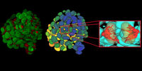
Transforming Research with Spatial Proteomics Workflows
Spatial Proteomics, Nature Methods 2024 Method of the Year, is driving research advancements in cancer, immunology, and beyond. By combining positional data with high throughput imaging of proteins in…
How Fluorescence Guides Sectioning of Resin-embedded EM Samples
Electron microscopes, including transmission electron microscopes (TEM) and scanning electron microscopes (SEM), are widely utilized to gain detailed structural information about biological samples or…
Coherent Raman Scattering Microscopy Publication List
CRS (Coherent Raman Scattering) microscopy is an umbrella term for label-free methods that image biological structures by exploiting the characteristic, intrinsic vibrational contrast of their…
Selecting the Right Dissecting Microscope
Learn how you can enhance dissection for life-science research and education with a microscope that ensures ergonomic comfort, high-quality optics, and easy access to the specimen.
Get to Insights Faster and Easier with AI Image Analysis Tools
Discover how Aivia helps scientists streamline image analysis with fast setup, accurate AI detection, and easy batch processing.
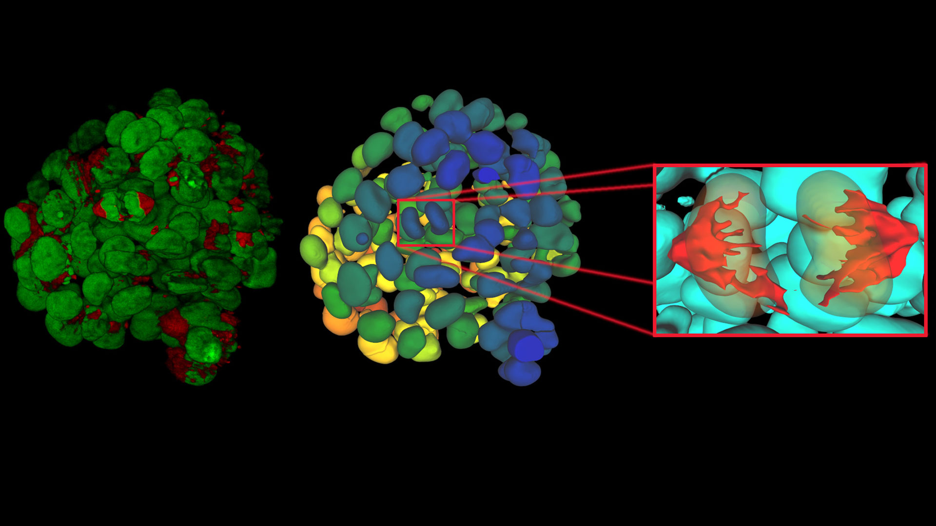
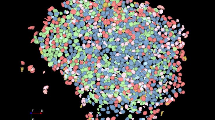
Unlocking the Secrets of Organoid Models in Biomedical Research
Get ready to delve deeper into the world of organoids and 3D models, which are essential tools for advancing our understanding of human health. Navigating these complex structures and obtaining clear…
ATTO-TEC Consumables
ATTO-TEC dyes have become a benchmark for fluorescence microscopy imaging, offering a highly differentiated panel. Their brightness and photostability make them the reagents of choice for demanding applications.

New platform with AI-powered search gives customers convenient access to tailored microscopy solutions
08 May 2025, Wetzlar, Germany - The Aivia 15 release includes deep learning-powered tools for 2D and 3D cell segmentation
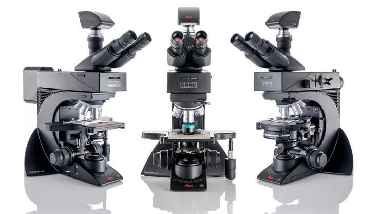
6 May 2025, Wetzlar, Germany - Leica Microsystems introduces the Visoria series to power up microscopy routines across industrial, life science and…
April 2025, Wetzlar, Germany – Leica Microsystems, a leading provider of microscopy and scientific information, has strengthened its partnership with…
