Leica DM3000 & DM3000 LED
Aufrechte Mikroskope
Lichtmikroskope
Produkte
Startseite
Leica Microsystems
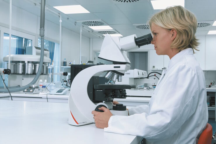
Leica DM3000 & DM3000 LED Systemmikroskope mit einzigartiger Ergonomie und intelligenter Automation:
Lesen Sie unsere neuesten Artikel
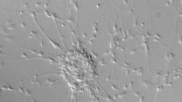
Differential Interference Contrast (DIC) Microscopy
This article demonstrates how differential interference contrast (DIC) can be actually better than brightfield illumination when using microscopy to image unstained biological specimens.
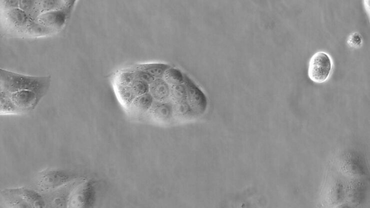
Phase Contrast and Microscopy
This article explains phase contrast, an optical microscopy technique, which reveals fine details of unstained, transparent specimens that are difficult to see with common brightfield illumination.
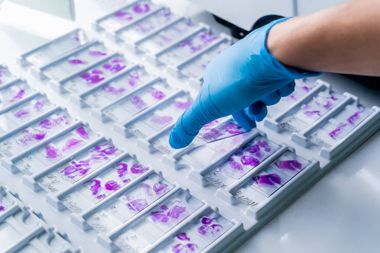
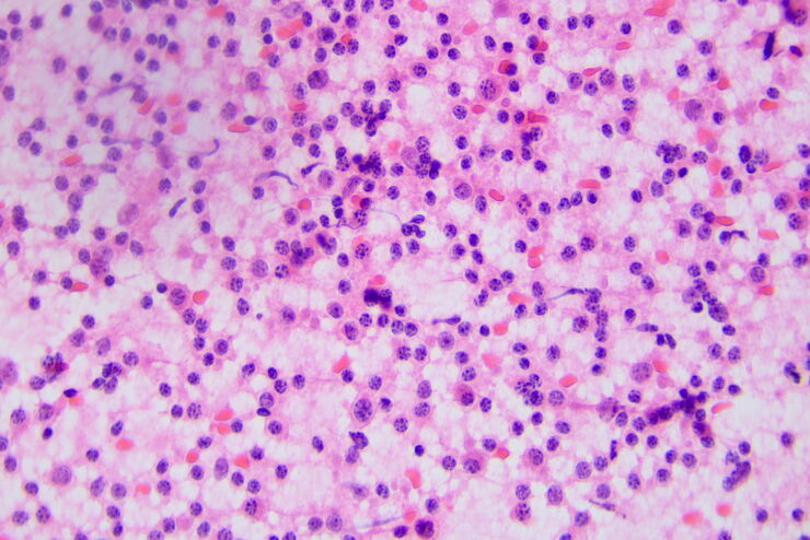
H&E Staining in Microscopy
If we consider the role of microscopy in pathologists’ daily routines, we often think of the diagnosis. While microscopes indeed play a crucial role at this stage of the pathology lab workflow, they…
How to Benefit from Digital Cytopathology
If you have thought of digital cytopathology as characterized by the digitization of glass slides, this webinar with Dr. Alessandro Caputo from the University Hospital of Salerno, Italy will broaden…
Factors to Consider when Selecting Clinical Microscopes
What matters if you would like to purchase a clinical microscope? Learn how to arrive at the best buying decision from our Science Lab Article.
The Time to Diagnosis is Crucial in Clinical Pathology
Abnormalities in tissues and fluids - that’s what pathologists are looking for when they examine specimens under the microscope. What they see and deduce from their findings is highly influential, as…
Perform Microscopy Analysis for Pathology Ergonomically and Efficiently
The main performance features of a microscope which are critical for rapid, ergonomic, and precise microscopic analysis of pathology specimens are described in this article. Microscopic analysis of…
Koehler Illumination: A Brief History and a Practical Set Up in Five Easy Steps
In this article, we will look at the history of the technique of Koehler Illumination in addition to how to adjust the components in five easy steps.
Anwendungsbereiche
Fluoreszenz
Die Fluoreszenz ist eines der am häufigsten verwendeten physikalischen Phänomene in der biologischen und analytischen Mikroskopie, vor allem wegen ihrer hohen Empfindlichkeit und Spezifität. Erfahren…
Klinische Pathologie
Erfahren Sie, wie ergonomische Mikroskope von Leica Microsystems eine genaue und zeitnahe Diagnose in der klinischen Pathologie unterstützen.
Mikroskopie in der Pathologie
Die pathologische Analyse von Proben erfordert manchmal lange Arbeitsstunden an einem Mikroskop. Dies kann für den Benutzer mit körperlichen Beschwerden und Belastungen verbunden sein, was zu einer…
Anatomische Pathologie
Erfahren Sie, wie ergonomische Mikroskope von Leica Microsystems eine effiziente und genaue Diagnose in der anatomischen Pathologie unterstützen.
DIC-Mikroskope
Ein DIC-Mikroskop ist ein Weitfeldmikroskop, bei dem sich zwischen Lichtquelle und Kondensorlinse sowie zwischen Objektiv und Kamerasensor oder Okularen ein Polarisationsfilter und ein…
Phasenkontrast-Lichtmikroskope
Mit einem Phasenkontrast-Lichtmikroskop können die Strukturen vieler Arten von biologischen Präparaten mit einem größeren Kontrast betrachtet werden, ohne dass die Probe eingefärbt werden muss.
Möchten Sie mehr erfahren?
Sprechen Sie mit unseren Experten.
Wünschen Sie eine persönliche Beratung? Show local contacts
