
Ciencias de la vida
Ciencias de la vida
Este es el lugar para ampliar sus conocimientos, capacidades de investigación y aplicaciones prácticas de la microscopía en diversos campos científicos. Aprenda a conseguir una visualización precisa, interpretación de imágenes y avances en la investigación. Encuentre información detallada sobre microscopía avanzada, técnicas de obtención de imágenes, preparación de muestras y análisis de imágenes. Los temas tratados incluyen la biología celular, la neurociencia y la investigación del cáncer, con especial atención a las aplicaciones e innovaciones de vanguardia.

How to Prepare your Specimen for Immunofluorescence Microscopy
Immunofluorescence (IF) specimen preparations can be analyzed by various fluorescence microscopy techniques, depending on the research application. IF is indispensable for researchers using a simple…

Live-Cell Imaging Techniques
The understanding of complex and/or fast cellular dynamics is an important step for exploring biological processes. Therefore, today’s life science research is increasingly focused on dynamic…

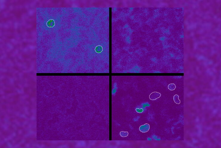
Imaging of Anti-Cancer Drug Uptake in Spheroids using DLS
Spheroid 3D cell culture models mimic the physiology and functions of living tissues making them a useful tool to study tumor morphology and screen anti-cancer drugs. The drug AZD2014 is a recognized…
 in 3D")
Artificial Intelligence and Confocal Microscopy – What You Need to Know
This list of frequently asked questions provides “hands-on” answers and is a supplement to the introductory article about Dynamic Signal Enhancement powered by Aivia "How Artificial Intelligence…
How Artificial Intelligence Enhances Confocal Imaging
In this article, we show how artificial intelligence (AI) can enhance your imaging experiments. Namely, how Dynamic Signal Enhancement powered by Aivia improves image quality while capturing the…

Benefits of Combining STED and Lifetime
In this interview, Professor Alberto Diaspro talks about the advantages of the White Light Laser and the TauSTED capabilities of STELLARIS 8 STED. He speaks about his experience with the confocal…
Spectroscopic Evaluation of Red Blood Cells
Hemoglobinopathies are a major healthcare problem. This study presents a possible diagnostic tool for thalassemia which is based on confocal spectroscopy. This approach exploits spectral detection and…

The Cryo-CLEM Journey
This article describes the Cryo-CLEM technology and the benefits it can provide for scientists. Additionally, some scientific publications are highlighted.
Recent developments in cryo electron…

Fluorescence Lifetime-based Imaging Gallery
Confocal microscopy relies on the effective excitation of fluorescence probes and the efficient collection of photons emitted from the fluorescence process. One aspect of fluorescence is the emission…
